Pancreatitis: Difference between revisions
No edit summary |
No edit summary |
||
| Line 107: | Line 107: | ||
* insulin injections, if the endocrine function of the pancreas is compromised | * insulin injections, if the endocrine function of the pancreas is compromised | ||
* analgesics (pain-relieving medication)<ref name=":1" /> | * analgesics (pain-relieving medication)<ref name=":1" /> | ||
== Physical Therapy Management == | == Physical Therapy Management == | ||
'''Acute Pancreatitis''' | '''Acute Pancreatitis''' | ||
Patients with acute pancreatitis may seek physical therapy treatment with a chief complaint of back pain. Back pain is common in patients with pancreatitis, as the inflammation and scarring associated with this disease can lead to decreased spinal extension, particularly in the thoracolumbar junction. This decrease in motion is difficult to treat even with patient compliance and resolution of inflammation due to the depth of the scarring. This tissue is often difficult to penetrate with mobilization techniques and, therefore, continues to decrease motion. Despite this, pain may be relieved through the use of heat to decrease muscular tension, relaxation techniques, and specific positioning techniques, including leaning forward, sitting up, or lying on the left side in the fetal position. | Patients with acute pancreatitis may seek physical therapy treatment with a chief complaint of [[Thoracic Back Pain|back pain]]. | ||
It is important to note that a patient may present to physical therapy prior to a diagnosis of acute pancreatitis with back pain. While acute pancreatitis is associated with gastrointestinal symptoms, including diarrhea, pain after eating, anorexia, and unexplained weight loss, the patient may not recognize the importance or necessity of reporting what they may believe are unrelated symptoms. Therefore physical therapists must thoroughly question patients about all body systems and warning signs. | |||
It is also important to know that pancreatitis is frequently associated with diabetes mellitus. Over 23 million Americans are currently living with diabetes, many of which will seek physical therapy services. Because of this it is important to be aware of the signs of symptoms of pancreatitis, as patients with diabetes are at an increased risk for this condition.<ref name="diabetes">American Diabetes Association. http://www.diabetes.org/ (accessed 11 April 2010).</ref> | * Back pain is common in patients with pancreatitis, as the inflammation and scarring associated with this disease can lead to decreased spinal extension, particularly in the thoracolumbar junction. This decrease in motion is difficult to treat even with patient compliance and resolution of inflammation due to the depth of the scarring. This tissue is often difficult to penetrate with [[Maitland's Mobilisations|mobilization]] techniques and, therefore, continues to decrease motion. Despite this, [[Pain Behaviours|pain]] may be relieved through the use of [[Heat Therapy|heat]] to decrease muscular tension, relaxation techniques, and specific positioning techniques, including leaning forward, sitting up, or lying on the left side in the fetal position. | ||
Patients with acute pancreatitis my also need physical therapy services if acute respiratory distress syndrome (ARDS) develops as a complication. Assisted respiration and pulmonary care are critical interventions for these patients. | * It is important to note that a patient may present to physical therapy prior to a diagnosis of acute pancreatitis with back pain. While acute pancreatitis is associated with gastrointestinal symptoms, including diarrhea, pain after eating, anorexia, and unexplained weight loss, the patient may not recognize the importance or necessity of reporting what they may believe are unrelated symptoms. Therefore physical therapists must thoroughly question patients about all body systems and warning signs. | ||
For patients who are receiving acute care and are restricted from eating or drinking in order to let the pancreas rest, even ice chips can stimulate enzymes and increase pain. Therefore, physical therapists must be careful to follow all medical orders and not adhere to patient requests until approved by the nursing or medical staff. | * It is also important to know that pancreatitis is frequently associated with [[Diabetes|diabetes mellitus]]. Over 23 million Americans are currently living with diabetes, many of which will seek physical therapy services. Because of this it is important to be aware of the signs of symptoms of pancreatitis, as patients with diabetes are at an increased risk for this condition.<ref name="diabetes">American Diabetes Association. http://www.diabetes.org/ (accessed 11 April 2010).</ref> | ||
Hospitalized patients with acute pancreatitis must also be monitored for signs and symptoms of bleeding, including bruising.<ref name="patho" /> | * Patients with acute pancreatitis my also need physical therapy services if acute respiratory distress syndrome ([[Acute Respiratory Distress Syndrome (ARDS)|ARDS]]) develops as a complication. Assisted respiration and pulmonary care are critical interventions for these patients. | ||
* For patients who are receiving acute care and are restricted from eating or drinking in order to let the pancreas rest, even ice chips can stimulate enzymes and increase pain. Therefore, physical therapists must be careful to follow all medical orders and not adhere to patient requests until approved by the nursing or medical staff. | |||
* Hospitalized patients with acute pancreatitis must also be monitored for signs and symptoms of bleeding, including bruising.<ref name="patho" /> | |||
'''''Chronic Pancreatitis''''' | '''''Chronic Pancreatitis''''' | ||
Similar to acute pancreatitis, patients with chronic pancreatitis may present to physical therapy with complaints of pain in the upper thoracic spine or at the thoracolumbar junction, while those with alcohol-related chronic pancreatitis may have symptoms of peripheral neuropathy. Because patients may complain of these seemingly musculoskeletal problems, it is imperative that physical therapists receive a complete history perform a thorough examination to screen for this disease. If this visceral problem is not determined upon initial evaluation, failure to improve with therapeutic intervention necessitates referral. | * Similar to acute pancreatitis, patients with chronic pancreatitis may present to physical therapy with complaints of pain in the upper thoracic spine or at the thoracolumbar junction, while those with alcohol-related chronic pancreatitis may have symptoms of peripheral [[Neuropathies|neuropathy]]. Because patients may complain of these seemingly musculoskeletal problems, it is imperative that physical therapists receive a complete history perform a thorough examination to screen for this disease. If this visceral problem is not determined upon initial evaluation, failure to improve with therapeutic intervention necessitates referral. | ||
Patients with known pancreatitis or post-pancreatectomy may need physical therapy services such as monitoring vital signs and/or blood glucose levels depending on what complications exist. Physical therapists should also educate these patients about the effects of malabsorption and associated osteoporosis that they may experience.<ref name="patho" /><br> | * Patients with known pancreatitis or post-pancreatectomy may need physical therapy services such as monitoring [[Vital Signs|vital signs]] and/or blood glucose levels depending on what complications exist. | ||
* Physical therapists should also educate these patients about the effects of malabsorption and associated [[osteoporosis]] that they may experience.<ref name="patho" /><br> | |||
== Differential Diagnosis == | == Differential Diagnosis == | ||
Revision as of 03:05, 3 September 2021
Original Editors - Amy Dean from Bellarmine University's Pathophysiology of Complex Patient Problems project. Top Contributors - Amy Dean, Admin, Lucinda hampton, George Prudden, WikiSysop, Kim Jackson, 127.0.0.1, Elaine Lonnemann, Dave Pariser, Wendy Walker and Scott Buxton
Introduction[edit | edit source]
Pancreatitis is inflammation of the pancreas, which can either be acute (sudden and severe) or chronic (ongoing). The pancreas is a gland that secretes both digestive enzymes and important hormones. Heavy alcohol consumption is one of the most common causes of chronic pancreatitis, followed by gallstones.
- Pancreatitis is one of the least common diseases of the digestive system. Treatment options include abstaining from alcohol, fasting until the inflammation subsides, medication and surgery[1].
- Pancreatitis is a potentially serious disorder characterized by inflammation of the pancreas that may cause autodigestion of the organ by its own enzymes. This disease has two manifestations: acute pancreatitis and chronic pancreatitis[2]
- Acute pancreatitis (AP) is an acute response to injury of the pancreas. Chronic pancreatitis (CP) can result in permanent damage to the structure and endocrine and exocrine functions of the pancreas.[3]
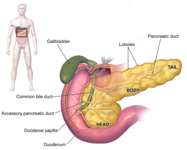
Image 1: Anatomy of the pancreas and its related organs, the gall bladder and duodenum
Acute Pancreatitis[edit | edit source]
Acute pancreatitis is the result of an inflammatory process involving the pancreas caused by the release of activated pancreatic enzymes.
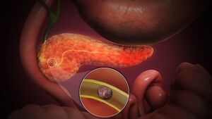
Image 2: 3D animation Acute pancreatitis
In addition to the pancreas, this disorder can also affect surrounding organs, as well as cause a systemic reaction. This form of pancreatitis is generally brief in duration, milder in symptom presentation, and reversible. However, while this form of the disease resolves both clinically and histologically, approximately 15% of patients with acute pancreatitis will develop chronic pancreatitis.[2][4] Acute pancreatitis may present as mild or severe. Milder forms of acute pancreatitis involve only the interstitium of the pancreas, which accounts for 80% of all cases, and has a temperate presentation with fewer complications. However, severe forms involve necrosis of the pancreatic tissue, which occurs in 20% of cases, and results in increased complications and mortality.[2][4]
Chronic Pancreatitis[edit | edit source]
Chronic pancreatitis develops from chronic inflammation of the pancreas that results in irreversible and progressive histologic changes. This includes fibrosis and ductal strictures, which destroy the pancreas directly, as well as decreased endocrine and exocrine functions, which can negatively affect other body systems. Unlike acute pancreatitis, this form of the disease is characterized by recurrent or persistent symptoms.[2][4]
Etiology[edit | edit source]
Around half of all people with acute pancreatitis have been heavy drinkers, which makes alcohol consumption one of the most common causes. Gallstones cause most of the remaining cases. In rare cases, pancreatitis can be caused by:
- trauma or surgery to the pancreas region
- inherited abnormalities of the pancreas
- inherited disorders of metabolism
- viruses (particularly mumps)
- medication (including some diuretics), which can also trigger inflammation[1].
Epidemiolgy[edit | edit source]
Acute pancreatitis accounts for about 275,000 hospital admissions annually.
- Eighty percent of patients admitted with pancreatitis usually have mild disease and can be discharged within a few days.
- Overall mortality of acute pancreatitis is approximately 2%.
- The relapse rate of acute pancreatitis is between 0.6% to 5.6%, and this depends on the etiology of pancreatitis. The relapse rate is highest when pancreatitis is due to alcohol use.
Chronic pancreatitis has an annual incidence rate of 5 to 12 per 100,000 people.
- The prevalence of chronic pancreatitis is 50 per 100,000 people.
- The most common age group is 30 to 40 years, and it occurs more in men than women[3].
Characteristics/Clinical Presentation[edit | edit source]
Abdominal pain is the most common presenting complaint of AP and can occur with nausea and vomiting. Chronic pancreatitis can present with or without abdominal pain, nausea or vomiting. Patients with chronic pancreatitis can present with steatorrhea and weight loss[3].
1.Common symptoms of an acute pancreatitis include: severe abdominal pain, often spreading through into the back; bloating; fever; sweating; nausea; vomiting; collapse.
2. Some people with chronic pancreatitis suffer recurrent or even constant abdominal pain, which may be severe. Other symptoms include steady weight loss, caused by the body’s inability to properly digest and absorb food. If much of the pancreas has been damaged, loss of insulin production can cause diabetes. Chronic pancreatitis can contribute to the development of pancreatic cancer.[3]
Associated Co-morbidities[edit | edit source]
Acute Pancreatitis
- Alcoholism
- 15% of patients with acute pancreatitis develop chronic pancreatitis
- 5-7% mortality rate for milder forms with inflammation confined to the pancreas
- 10-50% for severe forms with necrosis and hemorrhage of the gland and a systemic inflammatory response
- Infection of necrotic pancreatic tissue may occur after 5-7 days 100% mortality for pancreatic infection without extensive surgical debridement or drainage of the infected area
- Patients with peripancreatic inflammation or one area of fluid collection have a 10 to 15% chance of abscess formation
- Patients with two or more areas of fluid collection have a 60% incidence of abscess formation
- Diabetes mellitus (increased risk in alcoholic pancreatitis)
- Recurrent episodes (increased risk in alcoholic pancreatitis)[4][2]
Chronic Pancreatitis
- Alcoholism
- Cystic fibrosis
- Diabetes mellitus develops in 20-30% of patients within 10-15 years of onset
- Pancreatic cancer develops in 3% to 4% of patients
- Chronic disability
- 70% 10-year survival rate
- 45% 20-year survival rate
- 60% mortality rate for patients with alcohol-related chronic pancreatitis who do not cease alcohol intake[2]
Diagnosis[edit | edit source]
Pancreatitis is generally diagnosed quickly, by examination of the abdomen, and confirmed using a series of medical tests including:
- General tests – such as blood tests, physical examination and x-rays.
- Ultrasound – sound waves form a picture that detects the presence of gallstones.
- CT scan – a specialised x-ray takes three-dimensional pictures of the pancreas.
- MRI scan – this uses a strong magnetic field rather than radiation to take pictures of the abdomen. A special form of MRI called MRCP can also be used to get images of the ducts of the pancreas and help determine the cause of pancreatitis and the extent of damage[1].
Treatment[edit | edit source]
Treatment depends on the causes and severity of the condition.
Treatment for acute pancreatitis may include:
- hospital care – in all cases of acute pancreatitis
- intensive care in hospital – in cases of severe acute pancreatitis
- fasting and intravenous fluids – until the inflammation settles down
- pain relief – adequate pain relief is essential and is often given into the vein (intravenously). With appropriate pain relief, a person with pancreatitis is able to draw deep breaths, which helps to avoid lung complications such as pneumonia
- endoscopy – a thin tube is inserted through your oesophagus to allow the doctor to see your pancreas. This device is used to inject dye into the bile ducts and pancreas. Gallstones can be seen and removed directly
- surgery – if gallstones are present, removing the gallbladder will help prevent further attacks. In rare cases, surgery is needed to remove damaged or dead areas of the pancreas
- lifestyle change – not drinking alcohol.
Treatment for chronic pancreatitis may include:
- lowering fat intake
- supplementing digestion by taking pancreatic enzyme tablets with food
- cutting out alcohol
- insulin injections, if the endocrine function of the pancreas is compromised
- analgesics (pain-relieving medication)[1]
Physical Therapy Management[edit | edit source]
Acute Pancreatitis
Patients with acute pancreatitis may seek physical therapy treatment with a chief complaint of back pain.
- Back pain is common in patients with pancreatitis, as the inflammation and scarring associated with this disease can lead to decreased spinal extension, particularly in the thoracolumbar junction. This decrease in motion is difficult to treat even with patient compliance and resolution of inflammation due to the depth of the scarring. This tissue is often difficult to penetrate with mobilization techniques and, therefore, continues to decrease motion. Despite this, pain may be relieved through the use of heat to decrease muscular tension, relaxation techniques, and specific positioning techniques, including leaning forward, sitting up, or lying on the left side in the fetal position.
- It is important to note that a patient may present to physical therapy prior to a diagnosis of acute pancreatitis with back pain. While acute pancreatitis is associated with gastrointestinal symptoms, including diarrhea, pain after eating, anorexia, and unexplained weight loss, the patient may not recognize the importance or necessity of reporting what they may believe are unrelated symptoms. Therefore physical therapists must thoroughly question patients about all body systems and warning signs.
- It is also important to know that pancreatitis is frequently associated with diabetes mellitus. Over 23 million Americans are currently living with diabetes, many of which will seek physical therapy services. Because of this it is important to be aware of the signs of symptoms of pancreatitis, as patients with diabetes are at an increased risk for this condition.[5]
- Patients with acute pancreatitis my also need physical therapy services if acute respiratory distress syndrome (ARDS) develops as a complication. Assisted respiration and pulmonary care are critical interventions for these patients.
- For patients who are receiving acute care and are restricted from eating or drinking in order to let the pancreas rest, even ice chips can stimulate enzymes and increase pain. Therefore, physical therapists must be careful to follow all medical orders and not adhere to patient requests until approved by the nursing or medical staff.
- Hospitalized patients with acute pancreatitis must also be monitored for signs and symptoms of bleeding, including bruising.[2]
Chronic Pancreatitis
- Similar to acute pancreatitis, patients with chronic pancreatitis may present to physical therapy with complaints of pain in the upper thoracic spine or at the thoracolumbar junction, while those with alcohol-related chronic pancreatitis may have symptoms of peripheral neuropathy. Because patients may complain of these seemingly musculoskeletal problems, it is imperative that physical therapists receive a complete history perform a thorough examination to screen for this disease. If this visceral problem is not determined upon initial evaluation, failure to improve with therapeutic intervention necessitates referral.
- Patients with known pancreatitis or post-pancreatectomy may need physical therapy services such as monitoring vital signs and/or blood glucose levels depending on what complications exist.
- Physical therapists should also educate these patients about the effects of malabsorption and associated osteoporosis that they may experience.[2]
Differential Diagnosis[edit | edit source]
Acute Pancreatitis[edit | edit source]
Disorders presenting with symptoms similar to those of acute pancreatitis includeperforated gastric or duodenal ulcer, mesenteric infarction, medications, strangulating intestinal obstruction, dissecting aneurysm, biliary colic, appendicitis, diverticulitis, inferior wall myocardial infarction, tubo-ovarian abscess, renal failure, salivary gland disease, hematoma of the abdominal muscles or spleen, cholecystitis, vascular occlusions, pneumonia, hypertriglyceridemia, hypercalcemia, infection, post-traumatic injury, pregnancy, and diabetic ketoacidosis.[4][6][7][8]
Chronic Pancreatitis[edit | edit source]
Patients who do not present with a typical history of alcohol abuse and frequent episodes of acute pancreatitis, pancreatic malignancy must be ruled out as the cause of pain. In addition, chronic pancreatitis may initially be confused with acute pancreatitis because the symptoms are similar, gallstones, and neoplastic or inflammatory masses.[4][6]
Case Reports[edit | edit source]
Patient Demographics: Sixty-year-old male who was admitted to a hospital in October 2001
Chief Complaints: Abdominal pain and dark urine, which had begun 2 weeks previously
Past Medical History: Left lower extremity lymphedema, Raynauds disease, transient ischemic attacks, ischemic heart disease, left side hydronephrosis, lymph node biopsy in 1999, and chronic smoking
Laboratory Tests/Medical Imaging: Liver function tests, transabdominal ultrasound, ERCP, and a CT scan
Initial Medical Diagnosis: The patient was thought to have pancreatic cancer
Additional Tests: A biopsy was performed on a tumor found in the common bile duct, followed by palliative bypass surgery to decrease symptoms, undergo a gastrojejunostomy, choledochojejunostomy, and a cholecystectomy.These biopsies (pancreas, gallbladder, liver and the lymph node) were reviewed by a histopathologist and a diagnosis of sclerosing retroperitonitis was considered
Patient Symptom Progression: The patient developed a nodular itchy rash, with skin biopsies confirming this
Final Medical Diagnosis: Diagnosis of autoimmune pancreatitis
PT Relevance: This case illustrates how complex pancreatitis can present and how difficult it can be to diagnose, as several organs (pancreas, lymph node, liver, gallbladder, retroperitoneum, pericardium, and skin) can be involved at differed times during the disease process.
Nayar M, Charnley R, Scott J, Haugk B, and Oppong K. Autoimmune Pancreatitis with Multiorgan Involvement. A Case of Pericardial Involvement. Journal of the Pancreas. 2009; 10(5):539-542. http://www.joplink.net/. (accessed 27 March 2010).[9]
Patient Demographics: 57-year-old male presented
Chief Complaints: Acute epigastric pain, nausea, severe dehydration, and a dry mouth the morning after running a marathon. On the previous day, the patient had not only participated in a marathon, but also visited a sauna, and had inadequate fluid and food consumption during both events.
Physical Examination: The patient as found to have an expanded, hypertympanic and tender abdomen with active peristalsis
Vital Signs: Blood pressure of 165/100 mmHg, heart rate of 81 beats per minute, and a respiratory rate of 24 breaths/min
Laboratory Tests: Elevated glucose, amylase, CPK, and CRP levels
Medical imaging: Transabdominal ultrasonography found free intra-abdominal fluid containing blood and an amylase concentration of 944 U/L. Contrast enhanced CT scan showed that more than 90% of functioning pancreatic tissue was lost. 10 days after admission another CT scan showed necrotic pancreatic tissue and peripancreatic fluid collections
PT Relevance: This case is particularly useful for outpatient physical therapists, because while mechanical, stress, physical stress, or dehydration alone rarely cause damage, the combination of the combination of these factors can lead to pancreatic ischemia and, ultimately, acute pancreatitis.
Mast J, Morak M, Brett B, van Eijck C. Ischemic Acute Necrotizing Pancreatitis in a Marathon Runner. Journal of the Pancreas. 2009; 10(1):53-54. http://www.joplink.net/. (accessed 27 March 2010).[10]
Patient Demographics: 18-year-old man presented to the emergency room
Chief Complaints: Severe epigastric pain lasting 6 hours. Five hours prior to symptom onset, the patient ingested 7 tablets of 400 mg ibuprofen. He had also been taking ibuprofen as prescribed to treat low back pain for 1 week prior to this incident.
Laboratory Testing: Elevated serum amylase, urinary amylase, lactate dehydrogenase, and leucocytosis levels indicating acute pancreatitis
Medical Imaging: CT scan consistent with mild pancreatitis findings. To rule out the large dose of NSAID ingestion as the direct gastric source of epigastric pain a gastroscopy was performed, which was normal.
PT Relevance: While NSAIDs rarely induce acute pancreatic attacks, they, along with several other drugs, can cause acute pancreatitis. Because many patients seeking physical therapy treatment are also on some form of prescribed or over-the-counter pharmaceutical, it is important ask about medication use and watch for any related side effects. This article provides a list of Class I, II, and III drugs associated with acute pancreatitis that therapists can refer to.
Magill P, Ridgway P, Conlon K, Neary P. A Case of Probable Ibuprofen-Induced Acute Pancreatitis. Journal of the Pancreas. 2006; 7(3):311-314. http://www.joplink.net/. (accessed 21 March 2010).[11]
Patient Demographics: 16-year-old boy
Chief Complaints: Left paraumbilical, and occasionally epigastric, abdominal pain of moderate intensity. After 3 months of pain, these symptoms subsided. However, 20 days later the patient developed right-sided shoulder and chest pain, as well as dyspnea and sought medical treatment
Initial Medical Diagnosis: A physician misdiagnosed the patient’s problems as musculoskeletal pain
Patient Symptom Progression: As the patient’s symptoms of right-sided shoulder and chest pain and dyspnea persisted, he went to the hospital
Laboratory Tests: Elevated pleural amylase
Medical Imaging: Chest x-rays revealed a massive right-sided hemorrhagic pleural effusion and and abdominal CT scan showing a pancreatic pseudocyst. When the pleural effusion had not completely resolved following 3 weeks of treatment, another CT scan was performed, revealing that the pseudocyst was still present in the pancreas and causing the pleural effusion.
PT Relevance: This is important to note, as pancreatitis symptoms can manifest in many different ways and may be mistaken for pulmonary or musculoskeletal conditions if not tested and screened appropriately.
Namazi M and Mowla A. Massive right-sided hemorrhagic pleural effusion due to pancreatitis; a case report. BMC Pulmonary Medicine. 2004; 4:1. http://www.ncbi.nlm.nih.gov/pmc/articles/PMC362879/. (accessed 21 March 2010).[12]
Patient Demographics: 63-year-old female presented to the emergency department
Chief Complaint: Left flank and back pain persisting over the past 5 days. The patient had no fever, abdominal pain, chest pain, dyspnea, or symptoms related to the urinary system, nor had she suffered from any recent trauma.
Past Medical History: 5 year history of hypertension and type 2 diabetes mellitus that were being regularly treated, but no history of cardiac disease, stroke, renal disease, smoking, or alcohol consumption
Physical Examination: Left flank pain exacerbated with percussion
Laboratory Tests: Elevated C-reactive protein (CRP) levels; however, all other laboratory data, a urinary analysis, and abdominal x-rays were unremarkable
Medical Imaging: Due to the elevated CRP levels and left flank pain ultrasound was performed; however, there were no abnormal findings in the kidneys, spleen, pancreas, or hepatobiliary system. Because of this, a CT was performed which found abnormal fluid collection over the peri-renal space and pancreatic tail as well as necrotic changes and swelling of the pancreatic tail, while serum pancreatic enzymes revealed a normal amylase (90 u/L) and a slightly elevated lipase level (336 u/L)
Medical Diagnosis: Acute pancreatitis
PT Relevance: This case illustrates the fact that while pancreatitis typically manifests as upper abdominal pain, nausea, vomiting, and elevated amylase and lipase levels, patients may present with only one or a few of the typical symptoms. In this case, the only complain was left flank pain, a symptom that many physical therapy patients may present with. Therefore, it is important for physical therapists to investigate whether this pain is musculoskeletal or systemic in nature.
Chen JH, Chern CH, Chen JD, How CK, Wang LM, Lee CH. Left flank pain as the sole manifestation of acute pancreatitis: a report of a case with an initial misdiagnosis. Emerg Med J 2005;22:452-453. http://emj.bmj.com/content/22/6/452.full (accessed 31 March 2010).[13]
References[edit | edit source]
- ↑ 1.0 1.1 1.2 1.3 better health Pancreatitis Available:https://www.betterhealth.vic.gov.au/health/conditionsandtreatments/pancreatitis (accessed 3.9.2021)
- ↑ 2.0 2.1 2.2 2.3 2.4 2.5 2.6 2.7 Goodman CC, Fuller KS. Pathology: Implications for the Physical Therapist. 3rd ed. Saint Louis, MO: Saunders; 2009.
- ↑ 3.0 3.1 3.2 3.3 Mohy-ud-din N, Morrissey S. Pancreatitis.2019 Available: https://www.statpearls.com/articlelibrary/viewarticle/26577/(accessed 3.9.2021)
- ↑ 4.0 4.1 4.2 4.3 4.4 4.5 Beers MH, et. al. eds. The Merck Manual of Diagnosis and Therapy. 18th ed. Whitehouse Station, NJ: Merck Research Laboratories; 2006.
- ↑ American Diabetes Association. http://www.diabetes.org/ (accessed 11 April 2010).
- ↑ 6.0 6.1 Cleveland Clinic: Center for Continuing Education. Chronic Pancreatitis. http://www.clevelandclinicmeded.com/medicalpubs/diseasemanagement/gastroenterology/chronic-pancreatitis/ (accessed 21 March 2010).
- ↑ Carroll J, Herrick B, Gipson T. Acute Pancreatitis: Diagnosis, Prognosis, and Treatment. Am Fam Physician. 2008 Mar 1;77(5):594. http://www.aafp.org/afp/2007/0515/p1513.html (accessed 21 March 2010).
- ↑ The Department of Anaesthesia and Intensive Care. Acute Pancreatitis. http://www.aic.cuhk.edu.hk/web8/acute_pancreatitis.htm (accessed 21 March 2010).
- ↑ Nayar M, Charnley R, Scott J, Haugk B, and Oppong K. Autoimmune Pancreatitis with Multiorgan Involvement. A Case of Pericardial Involvement. Journal of the Pancreas. 2009; 10(5):539-542. http://www.joplink.net/. (accessed 27 March 2010).
- ↑ Mast J, Morak M, Brett B, van Eijck C. Ischemic Acute Necrotizing Pancreatitis in a Marathon Runner. Journal of the Pancreas. 2009; 10(1):53-54. http://www.joplink.net/. (accessed 27 March 2010).
- ↑ Magill P, Ridgway P, Conlon K, Neary P. A Case of Probable Ibuprofen-Induced Acute Pancreatitis. Journal of the Pancreas. 2006; 7(3):311-314. http://www.joplink.net/. (accessed 21 March 2010).
- ↑ Namazi M and Mowla A. Massive right-sided hemorrhagic pleural effusion due to pancreatitis; a case report. BMC Pulmonary Medicine. 2004; 4:1. http://www.ncbi.nlm.nih.gov/pmc/articles/PMC362879/. (accessed 21 March 2010).
- ↑ Chen JH, Chern CH, Chen JD, How CK, Wang LM, Lee CH. Left flank pain as the sole manifestation of acute pancreatitis: a report of a case with an initial misdiagnosis. Emerg Med J 2005;22:452-453. http://emj.bmj.com/content/22/6/452.full (accessed 31 March 2010).


