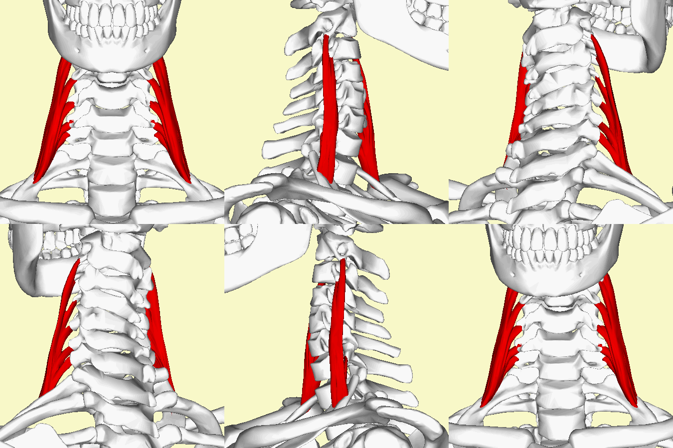
Middle Scalene
Original Editor - Wendy Walker
Lead Editors - Wendy Walker, Kim Jackson, Milijana Delevic, Admin, WikiSysop, Wendy Snyders and Evan Thomas
Description[edit | edit source]
Middle Scalene, AKA Scalenus Medius (Latin: musculus scalenus medius), is the largest and longest muscle in the scalene group of lateral neck muscles. Often penetrated by the dorsal scapular and long thoracic nerves, it is deeply placed, lying behind Sternocleidomastoid.
Origin[edit | edit source]
Insertion[edit | edit source]
Upper surface of the 1st rib, between the scalene tubercle and the subclavian groove
Nerve Supply[edit | edit source]
Anterior rami of cervical spinal nerves C3 - C7
Blood Supply[edit | edit source]
Muscular branches of the ascending cervical artery
Action[edit | edit source]
Middle scalene can act with anterior and posterior scalene muscles or individually:[1]
- When scalenes act together - neck flexes. Also, they elevate 1st rib (and thorax) during a strong inhalation which is why they are called accessory muscles of inspiration.
- Individually - cervical lateral flexion and rotation[2]
Clinically important anatomy[edit | edit source]
Interscalene triangle [3] is the anatomical triangle formed between the scalenus anterior muscle, the scalenus medius muscle and the first rib. The space is very narrow and some structures like subclavian artery and brachial plexus pass through. When scalenes get tight, the narrow space becomes narrower and a person can develop pain, paresthesia and/or circulatory problems - Scalene syndrome or Thoracic Outlet Syndrome (TOS)
References[edit | edit source]
- ↑ https://www.nielasher.com/blogs/video-blog/trigger-point-therapy-treating-the-scalenes
- ↑ J Orthop Sports Phys Ther. 2002 Oct;32(10):488-96.Actions of the scalene muscles for rotation of the cervical spine in macaque and human. Buford JA, Yoder SM, Heiss DG, Chidley JV.
- ↑ https://www.kenhub.com/en/library/anatomy/scalene-muscles


