Hangman's Fracture: Difference between revisions
No edit summary |
Kim Jackson (talk | contribs) No edit summary |
||
| (33 intermediate revisions by 11 users not shown) | |||
| Line 4: | Line 4: | ||
'''Top Contributors''' - {{Special:Contributors/{{FULLPAGENAME}}}} | '''Top Contributors''' - {{Special:Contributors/{{FULLPAGENAME}}}} | ||
<br></div> | <br></div> | ||
== | == Definition/Description == | ||
Hangman’s fracture is | Hangman’s fracture also known as Traumatic Spondylolisthesis of Axis is defined as bilateral fracture traversing the pars interarticularis of cervical vertebrae 2 (C2) with an associated traumatic subluxation of C2 on cervical vertebrae 3 (C3). It is the second most common fracture of the C2 vertebrae following a fracture of the odontoid process. <ref name=":0" /><ref name=":1">Al-Mahfoudh R, Beagrie C, Woolley E, Zakaria R, Radon M, Clark S, Pillay R, Wilby M. [https://www.ncbi.nlm.nih.gov/pmc/articles/PMC4836940/ Management of typical and atypical Hangman's fractures.] Global spine journal. 2016 May;6(03):248-56.</ref> | ||
== Clinically Relevant Anatomy | == Clinically Relevant Anatomy == | ||
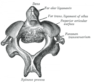
<u></u>A hangman’s fracture is the result of hyperextension of the upper cervical spine. | [[Image:Axis.png|right|300px|Axis - C2]]<u></u>A hangman’s fracture is the result of hyperextension of the upper cervical spine. In '''typical''' hangman's fracture the pedicles of the axis break symmetrically. While in '''atypical''' hangman's fracture, the fracture is in the posterior part of the vertebral body, on one or both sides. In contrary to what everyone thinks, the dens of C2 always remains intact. The pedicles are the smallest parts of the bony ring of the axis. They are additional weakened by the foramen transversarium on each side. There is no other cervical bony injury with a Hangman’s fracture.<ref name=":0">T.G. Williams. Hangman’s Fracture. The journal of bone and Joint surgery, pp. 82-89</ref><ref name=":1" /> | ||
Other tissues that may be damaged are the anterior ligament and the disc below the axis, both may be disrupted. Neurological injuries are rare because the spinal canal is sufficiently wide at this level. | Other tissues that may be damaged are the anterior ligament and the disc below the axis, both may be disrupted. Neurological injuries are rare because the spinal canal is sufficiently wide at this level. | ||
== Mechanism of Injury | == Etiology/Mechanism of Injury == | ||
The mechanism of injury is a forced hyperextension of the head with distraction of the neck. This occurs when the nose is placed under the subject's chin such as in the case of a judicial hanging. | |||
The more common mechanism of action is hyperextension and axial loading. These injuries are most commonly seen in motor vehicle accidents, diving injuries, or contact sports.. Hangman’s fractures also occur in car accidents when an unrestrained passenger or driver strikes the dashboard or when rebound hyperextension en distraction of the neck.<ref>Yilmaz, F., Akbulut, A. & Kose, O. (2010). An unusual presentation of an atypical hangman’s fracture. J Emerg Trauma Shock, 3(3), pp. 292–293.</ref> | |||
In elderly people, a hangman's fracture can be caused by low-impact trauma as well. This is more common in people who have conditions like osteoporosis, cancer that has spread to the bones, or vitamin D deficiency. | |||
== Clinical Presentation == | == Clinical Presentation == | ||
* Non-specific suboccipital pain. | |||
* Spasm of the neck muscles. | |||
* Numbness | |||
* Bruising around the neck | |||
* Troubled breathing<ref>Pathria, M.N. Physical injury: Spine. In: Resnick D, ed. Diagnosis of Bone and Joint Disorders. W.B. Saunders Co.; 2002; 2964-2967</ref>. | |||
== Diagnostic Procedures == | |||
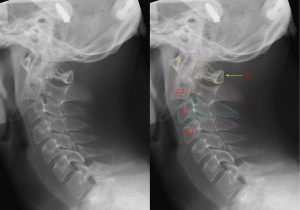
[[Image:Hangman's fracture.JPG|right|300x300px|X-ray of the cervical spine with a Hangman's fracture. Left without, right with annotation. C2 (outlined in red) is moved forward with respect to C3 (outlined in blue).|frameless]] | |||
The diagnosis of a hangman’s fracture can be made using x- ray and CT scan. MRI can be done to check for ligamentous and/or soft tissue injury. | |||
== Classification == | |||
Multiple grading systems for hangman’s fractures exist; however, Effendi<ref>Effendi, B., Roy, D., Cornish B, Dussault, R.G. & Laurin, C.A. (1981). Fractures of the ring of the axis: a classification based on analysis or 131 cases. J Bone Joint Surg Br, 63, pp. 319–27.</ref>and the Levine and Edwards <ref name="Li">Li, X., Dai, L., Lu, H. & Chen X. (2006). [https://link.springer.com/article/10.1007/s00586-005-0918-2 A systematic review of the management of hangman's fractures]. European Spine Journal, 15(3), pp. 257-269.</ref>classifications are the most widely used. | |||
#Type I: Caused by axial loading and hyperextension. There is | Levine and Edwards classification: | ||
#Type I: Caused by axial loading and hyperextension. There is less than 3mm subluxation of C2 on C3 and no angulation. This fracture is stable because the anterior longitudinal ligament and posterior longitudinal ligaments are intact along with the C2/C3 disk. | |||
#Type II: Caused by axial loading and hyperextension followed by a rebound flexion and axial loading. There is subluxation of C2 on C3 greater than 4mm or 11° angulation. There is some damage to the anterior longitudinal ligament and significant structural damage to the posterior longitudinal ligament and C2/C3 disk. This fracture is unstable. | #Type II: Caused by axial loading and hyperextension followed by a rebound flexion and axial loading. There is subluxation of C2 on C3 greater than 4mm or 11° angulation. There is some damage to the anterior longitudinal ligament and significant structural damage to the posterior longitudinal ligament and C2/C3 disk. This fracture is unstable. | ||
#Type IIA: Caused by flexion with distraction. These have no anterior displacement, but there is a severe angulation. The C2/C3 disk is damaged along with the posterior longitudinal ligaments. This fracture is unstable | #Type IIA: Caused by flexion with distraction. These have no anterior displacement, but there is a severe angulation. The C2/C3 disk is damaged along with the posterior longitudinal ligaments. This fracture is unstable | ||
#Type III: Caused by initial flexion and rebound extension with axial compression. There is severe displacement and angulation. The injury is accompanied by C2/C3 disk dislocation and unilateral or bilateral C2/C3 facet dislocation. There is also injury of the anterior and posterior longitudinal ligament. This fracture is considered unstable. | #Type III: Caused by initial flexion and rebound extension with axial compression. There is severe displacement and angulation. The injury is accompanied by C2/C3 disk dislocation and unilateral or bilateral C2/C3 facet dislocation. There is also injury of the anterior and posterior longitudinal ligament. This fracture is considered unstable. | ||
The | == Outcome Measures == | ||
The healing rate can be evaluated by radiological appearance. The same system of Levine-Edwards is used for evaluating. Type I hangman’s fracture has a healing rate of almost 100%. The healing rate drops as the fracture becomes more unstable<ref name="Li" />. The ‘Neck Disability Index (NDI)’ can be used to evaluate the effects of treatment<ref>Vernon, H. & Mior, S. (1991). The Neck Disability Index: a study of reliability an validity. Journal of Manipulative and Physiological Therapeutics, 14(7), pp. 409-415.</ref>. | |||
== | == Management / Interventions == | ||
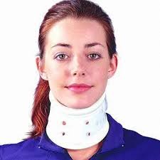
Treatment options are usually based on the stability of the hangman’s fracture. But most publications advocate that the primary treatment should be conservative.<ref name="Li" />Surgical interventions like internal fixation are usually performed in unstable fractures or when there is a possible risk of later instability. [[File:Rigid collar 1.jpg|thumb|Rigid cervical collar]] | |||
The physiotherapeutic intervention is a conservative treatment consisting of a period of traction and external immobilization. The external immobilization can be rigid or nonrigid depending on the type of fracture. The conservative treatment has more chance of being effective for stable and neurologically normal patients. | |||
<br>Effendi type I, II and Levine-Edwards type II fractures can be treated with nonrigid immobilization alone. | |||
Type IIa and III Levine-Edwards fractures are to be treated with rigid immobilization and traction<ref name="Li" />. In case of significant dislocation, fracture needs to be treated surgically. | |||
Cervical collar immobilization can be used for the displacements up to 6mm.<ref name=":1" /><br>The use of the immobilization brace (Halo-Vest) should be well considered and explained to the patient. A recent publication reported a 39,1% failure rate and 60,9% chance on complications<ref>Shin, J.J., Kim, S.J., Kim, T.H., Shin, H.S., Hwang, Y.S. & Park, S.K. (2010). Optimal use of the halo-vest orthosis for upper cercival spine injuries. Yonsei Medical Journal, 51(5), pp. 648-652.</ref>. It is recommended for type IIa and III fractures.<ref name=":1" /> | |||
Atypical hangman's fracture has more instability, so it requires surgical fixation.<ref name=":1" /> | |||
Since literature is scare the optimal treatment is not yet clear. | Since literature is scare the optimal treatment is not yet clear. | ||
| Line 49: | Line 66: | ||
== Differential Diagnosis == | == Differential Diagnosis == | ||
Differential diagnoses include pseudo-subluxation (generally C2 on C3) and the Mach effect.<ref name=":2" /> | |||
== Complications == | |||
* Vertebral artery arteriovenous fistula (AVF). | |||
* Occlusion and dissection of the vertebral artery. | |||
* Brown-Sequard syndrome (BSS). | |||
* Concurrent spinal cord injury especially in atypical, Type IIa, and III fractures.<ref name=":2">''StatPearls'': "Anatomy, Head and Neck, Cervical Vertebrae," "Hangman's Fractures."</ref> | |||
== References == | == References == | ||
<references /> | <references /> | ||
[[Category:Cervical Spine]] | |||
[[Category: | [[Category:Cervical Spine - Conditions]] | ||
[[Category:Conditions]] | |||
[[Category:Fractures]] | |||
[[Category:Primary Contact]] | |||
[[Category:Acute Care]] | |||
Latest revision as of 18:59, 19 November 2023
Original Editor - Thomas Maeseele
Top Contributors - Tarina van der Stockt, Rachael Lowe, Kim Jackson, Riya Naval, Thomas Maeseele, Lilian Ashraf, Sehriban Ozmen, 127.0.0.1, Admin, Evan Thomas, WikiSysop, Karen Wilson and Claire Knott
Definition/Description[edit | edit source]
Hangman’s fracture also known as Traumatic Spondylolisthesis of Axis is defined as bilateral fracture traversing the pars interarticularis of cervical vertebrae 2 (C2) with an associated traumatic subluxation of C2 on cervical vertebrae 3 (C3). It is the second most common fracture of the C2 vertebrae following a fracture of the odontoid process. [1][2]
Clinically Relevant Anatomy[edit | edit source]
A hangman’s fracture is the result of hyperextension of the upper cervical spine. In typical hangman's fracture the pedicles of the axis break symmetrically. While in atypical hangman's fracture, the fracture is in the posterior part of the vertebral body, on one or both sides. In contrary to what everyone thinks, the dens of C2 always remains intact. The pedicles are the smallest parts of the bony ring of the axis. They are additional weakened by the foramen transversarium on each side. There is no other cervical bony injury with a Hangman’s fracture.[1][2]
Other tissues that may be damaged are the anterior ligament and the disc below the axis, both may be disrupted. Neurological injuries are rare because the spinal canal is sufficiently wide at this level.
Etiology/Mechanism of Injury[edit | edit source]
The mechanism of injury is a forced hyperextension of the head with distraction of the neck. This occurs when the nose is placed under the subject's chin such as in the case of a judicial hanging.
The more common mechanism of action is hyperextension and axial loading. These injuries are most commonly seen in motor vehicle accidents, diving injuries, or contact sports.. Hangman’s fractures also occur in car accidents when an unrestrained passenger or driver strikes the dashboard or when rebound hyperextension en distraction of the neck.[3]
In elderly people, a hangman's fracture can be caused by low-impact trauma as well. This is more common in people who have conditions like osteoporosis, cancer that has spread to the bones, or vitamin D deficiency.
Clinical Presentation[edit | edit source]
- Non-specific suboccipital pain.
- Spasm of the neck muscles.
- Numbness
- Bruising around the neck
- Troubled breathing[4].
Diagnostic Procedures[edit | edit source]
The diagnosis of a hangman’s fracture can be made using x- ray and CT scan. MRI can be done to check for ligamentous and/or soft tissue injury.
Classification[edit | edit source]
Multiple grading systems for hangman’s fractures exist; however, Effendi[5]and the Levine and Edwards [6]classifications are the most widely used.
Levine and Edwards classification:
- Type I: Caused by axial loading and hyperextension. There is less than 3mm subluxation of C2 on C3 and no angulation. This fracture is stable because the anterior longitudinal ligament and posterior longitudinal ligaments are intact along with the C2/C3 disk.
- Type II: Caused by axial loading and hyperextension followed by a rebound flexion and axial loading. There is subluxation of C2 on C3 greater than 4mm or 11° angulation. There is some damage to the anterior longitudinal ligament and significant structural damage to the posterior longitudinal ligament and C2/C3 disk. This fracture is unstable.
- Type IIA: Caused by flexion with distraction. These have no anterior displacement, but there is a severe angulation. The C2/C3 disk is damaged along with the posterior longitudinal ligaments. This fracture is unstable
- Type III: Caused by initial flexion and rebound extension with axial compression. There is severe displacement and angulation. The injury is accompanied by C2/C3 disk dislocation and unilateral or bilateral C2/C3 facet dislocation. There is also injury of the anterior and posterior longitudinal ligament. This fracture is considered unstable.
Outcome Measures[edit | edit source]
The healing rate can be evaluated by radiological appearance. The same system of Levine-Edwards is used for evaluating. Type I hangman’s fracture has a healing rate of almost 100%. The healing rate drops as the fracture becomes more unstable[6]. The ‘Neck Disability Index (NDI)’ can be used to evaluate the effects of treatment[7].
Management / Interventions[edit | edit source]
Treatment options are usually based on the stability of the hangman’s fracture. But most publications advocate that the primary treatment should be conservative.[6]Surgical interventions like internal fixation are usually performed in unstable fractures or when there is a possible risk of later instability.
The physiotherapeutic intervention is a conservative treatment consisting of a period of traction and external immobilization. The external immobilization can be rigid or nonrigid depending on the type of fracture. The conservative treatment has more chance of being effective for stable and neurologically normal patients.
Effendi type I, II and Levine-Edwards type II fractures can be treated with nonrigid immobilization alone.
Type IIa and III Levine-Edwards fractures are to be treated with rigid immobilization and traction[6]. In case of significant dislocation, fracture needs to be treated surgically.
Cervical collar immobilization can be used for the displacements up to 6mm.[2]
The use of the immobilization brace (Halo-Vest) should be well considered and explained to the patient. A recent publication reported a 39,1% failure rate and 60,9% chance on complications[8]. It is recommended for type IIa and III fractures.[2]
Atypical hangman's fracture has more instability, so it requires surgical fixation.[2]
Since literature is scare the optimal treatment is not yet clear.
Differential Diagnosis[edit | edit source]
Differential diagnoses include pseudo-subluxation (generally C2 on C3) and the Mach effect.[9]
Complications[edit | edit source]
- Vertebral artery arteriovenous fistula (AVF).
- Occlusion and dissection of the vertebral artery.
- Brown-Sequard syndrome (BSS).
- Concurrent spinal cord injury especially in atypical, Type IIa, and III fractures.[9]
References[edit | edit source]
- ↑ 1.0 1.1 T.G. Williams. Hangman’s Fracture. The journal of bone and Joint surgery, pp. 82-89
- ↑ 2.0 2.1 2.2 2.3 2.4 Al-Mahfoudh R, Beagrie C, Woolley E, Zakaria R, Radon M, Clark S, Pillay R, Wilby M. Management of typical and atypical Hangman's fractures. Global spine journal. 2016 May;6(03):248-56.
- ↑ Yilmaz, F., Akbulut, A. & Kose, O. (2010). An unusual presentation of an atypical hangman’s fracture. J Emerg Trauma Shock, 3(3), pp. 292–293.
- ↑ Pathria, M.N. Physical injury: Spine. In: Resnick D, ed. Diagnosis of Bone and Joint Disorders. W.B. Saunders Co.; 2002; 2964-2967
- ↑ Effendi, B., Roy, D., Cornish B, Dussault, R.G. & Laurin, C.A. (1981). Fractures of the ring of the axis: a classification based on analysis or 131 cases. J Bone Joint Surg Br, 63, pp. 319–27.
- ↑ 6.0 6.1 6.2 6.3 Li, X., Dai, L., Lu, H. & Chen X. (2006). A systematic review of the management of hangman's fractures. European Spine Journal, 15(3), pp. 257-269.
- ↑ Vernon, H. & Mior, S. (1991). The Neck Disability Index: a study of reliability an validity. Journal of Manipulative and Physiological Therapeutics, 14(7), pp. 409-415.
- ↑ Shin, J.J., Kim, S.J., Kim, T.H., Shin, H.S., Hwang, Y.S. & Park, S.K. (2010). Optimal use of the halo-vest orthosis for upper cercival spine injuries. Yonsei Medical Journal, 51(5), pp. 648-652.
- ↑ 9.0 9.1 StatPearls: "Anatomy, Head and Neck, Cervical Vertebrae," "Hangman's Fractures."


