Gluteus Maximus: Difference between revisions
No edit summary |
No edit summary |
||
| Line 8: | Line 8: | ||
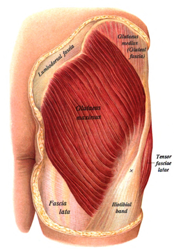
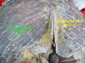
[[Image:Glutmax2.png|frame|right|Gluteus Maximus]] The largest of all gluteal muscles that is located at the posterior aspect of hip joint. Its size allows it to generate a large amount of force. The muscle evolved from an adductor of the hip which is still seen in lower primates today. The development of the muscle's function is associated with the erect posture and changes to the pelvis. It now functions to maintain the erect posture as one of the muscles that extends the hip joint.<br> | [[Image:Glutmax2.png|frame|right|Gluteus Maximus]] The largest of all gluteal muscles that is located at the posterior aspect of hip joint. Its size allows it to generate a large amount of force. The muscle evolved from an adductor of the hip which is still seen in lower primates today. The development of the muscle's function is associated with the erect posture and changes to the pelvis. It now functions to maintain the erect posture as one of the muscles that extends the hip joint.<br> | ||
The fibres of Gluteal maximus are largely perpendicular to each other and line up in the direction of pull giving it it's quadrilateral shape and course appearance. There are two layers to the muscle which pass down to the insertional attachment.<ref name="pala">Palastanga N, Soames R. Anatomy and Human Movement: Structure and Function. 6th ed. London, United Kingdom: Churchill Livingstone; 2012.</ref> | The fibres of Gluteal maximus are largely perpendicular to each other and line up in the direction of pull giving it it's quadrilateral shape and course appearance. There are two layers to the muscle which pass down to the insertional attachment.<ref name="pala">Palastanga N, Soames R. Anatomy and Human Movement: Structure and Function. 6th ed. London, United Kingdom: Churchill Livingstone; 2012.</ref> | ||
= Anatomy = | = Anatomy = | ||
| Line 22: | Line 22: | ||
=== Insertion === | === Insertion === | ||
Three-quarters of the fibres form a separate superficial lamnina which narrows and attaches between the two layers of the [[ | Three-quarters of the fibres form a separate superficial lamnina which narrows and attaches between the two layers of the [[Tensor Fascia Lata|tensor facscia lata]], thereby helping to form the '''iliotibial tract'''. | ||
The deeper fibres form an aponeurosis which attaches to the '''gluteal tuberosity''' of the femur.<ref name="pala" /> | The deeper fibres form an aponeurosis which attaches to the '''gluteal tuberosity''' of the femur.<ref name="pala" /> | ||
=== Nerve supply === | === Nerve supply === | ||
The gluteus maximus is supplied by the inferior gluteal nerve (root L5, S1 and S2). Cutaneous supply is mainly provided by L2 and 3.<ref name="pala" /> | The gluteus maximus is supplied by the inferior gluteal nerve (root L5, S1 and S2). Cutaneous supply is mainly provided by L2 and 3.<ref name="pala" /> | ||
= Function = | = Function = | ||
| Line 36: | Line 36: | ||
As a powerful extensor of the hip joint, the gluteus maximus is suited to powerful lower limb movements such as stepping onto a step, climbing or running but is not used greatly during normal walking. | As a powerful extensor of the hip joint, the gluteus maximus is suited to powerful lower limb movements such as stepping onto a step, climbing or running but is not used greatly during normal walking. | ||
Research has indicated that contraction of the deep abdominal muscles may assist with the contraction of gluteus maximus to assist with the control of anterior pelvic rotation.<ref name="kim">Kim TW, Kim YW.Effects of abdominal drawing-in during prone hip extension on the muscle activities of the hamstring, gluteus maximus, and lumbar erector spinae in subjects with lumbar hyperlordosis; J Phys Ther Sci. 2015 Feb;27(2):383-6</ref> Gluteal muscle weakness has been proposed to be associated with a number of lower limb injuries.<ref name=" | Research has indicated that contraction of the deep abdominal muscles may assist with the contraction of gluteus maximus to assist with the control of anterior pelvic rotation.<ref name="kim">Kim TW, Kim YW.Effects of abdominal drawing-in during prone hip extension on the muscle activities of the hamstring, gluteus maximus, and lumbar erector spinae in subjects with lumbar hyperlordosis; J Phys Ther Sci. 2015 Feb;27(2):383-6</ref> Gluteal muscle weakness has been proposed to be associated with a number of lower limb injuries.<ref name="dist">Distefano LJ, Blackburn JT, Marshall SW, Padua DAGluteal muscle activation during common therapeutic exercises; J Orthop Sports Phys Ther. 2009 Jul;39(7):532-40</ref> | ||
Effective exercise to specifically target the gluteus maximus muscle includes the single leg squat and the singe leg prone dead lift. These exercises elicited the most significant activity in electromyography(EMG) when tested against other forms of exercise. | Effective exercise to specifically target the gluteus maximus muscle includes the single leg squat and the singe leg prone dead lift. These exercises elicited the most significant activity in electromyography(EMG) when tested against other forms of exercise.<ref name="dist" /> | ||
= Assessment = | = Assessment = | ||
| Line 72: | Line 72: | ||
= See also = | = See also = | ||
*[[ | *[[Gluteus Medius|Gluteus medius]]<br> | ||
*[[ | *[[Lower crossed syndrome|Lower crossed syndrome]]<br> | ||
*[[Ober' | *[[Ober's Test|Ober's test]] | ||
= Recent Related Research (from Pubmed) = | = Recent Related Research (from Pubmed) = | ||
<div class="researchbox"><rss>http://www.ncbi.nlm.nih.gov/entrez/eutils/erss.cgi?rss_guid=1X9MO_201KJBQIff2kV1pqxLVbLESAHxiZ-c8SF7TQx_44T5p2|charset=UTF-8|short|max=10</rss></div> | <div class="researchbox"><rss>http://www.ncbi.nlm.nih.gov/entrez/eutils/erss.cgi?rss_guid=1X9MO_201KJBQIff2kV1pqxLVbLESAHxiZ-c8SF7TQx_44T5p2|charset=UTF-8|short|max=10</rss></div> | ||
= References = | = References = | ||
Revision as of 17:30, 22 February 2016
Original Editor - Joanne Garvey
Top Contributors - George Prudden, Joanne Garvey, Kim Jackson, Ahmed Nasr, Lucinda hampton, Admin, Joao Costa, Rachael Lowe, Khloud Shreif, Richard Benes, Wendy Walker, Evan Thomas, WikiSysop, Sai Kripa, Kai A. Sigel and Candace Goh
Description[edit | edit source]
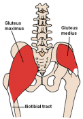
The largest of all gluteal muscles that is located at the posterior aspect of hip joint. Its size allows it to generate a large amount of force. The muscle evolved from an adductor of the hip which is still seen in lower primates today. The development of the muscle's function is associated with the erect posture and changes to the pelvis. It now functions to maintain the erect posture as one of the muscles that extends the hip joint.
The fibres of Gluteal maximus are largely perpendicular to each other and line up in the direction of pull giving it it's quadrilateral shape and course appearance. There are two layers to the muscle which pass down to the insertional attachment.[1]
Anatomy[edit | edit source]
Origin[edit | edit source]
Gluteal surface of illium behind the posterior gluteal line, posterior border of the illium, and the adjacent part of the iliac crest
Additionally, the side of the coccyx and posterior aspect of the sacrum.
Fibres also attach to the upper sacrotuberous ligament and aponeurosis of the sacrospinalis.[1]
Insertion[edit | edit source]
Three-quarters of the fibres form a separate superficial lamnina which narrows and attaches between the two layers of the tensor facscia lata, thereby helping to form the iliotibial tract.
The deeper fibres form an aponeurosis which attaches to the gluteal tuberosity of the femur.[1]
Nerve supply[edit | edit source]
The gluteus maximus is supplied by the inferior gluteal nerve (root L5, S1 and S2). Cutaneous supply is mainly provided by L2 and 3.[1]
Function[edit | edit source]
Gluteus maximus acts to extend and laterally rotate the hip joint. Futhermore upper fibres can abduct whereas the lower fibres can adduct the hip. Superior fibres can extend the knee through its attachment to the iliotibial tract as well as providing support to lateral knee. When the femur is fixed gluteus maximus pulls the pevis backwards causing the trunk to extend from a flexed position.
As a powerful extensor of the hip joint, the gluteus maximus is suited to powerful lower limb movements such as stepping onto a step, climbing or running but is not used greatly during normal walking.
Research has indicated that contraction of the deep abdominal muscles may assist with the contraction of gluteus maximus to assist with the control of anterior pelvic rotation.[2] Gluteal muscle weakness has been proposed to be associated with a number of lower limb injuries.[3]
Effective exercise to specifically target the gluteus maximus muscle includes the single leg squat and the singe leg prone dead lift. These exercises elicited the most significant activity in electromyography(EMG) when tested against other forms of exercise.[3]
Assessment[edit | edit source]
Palpation[edit | edit source]
Power[edit | edit source]
Length[edit | edit source]
Treatment[edit | edit source]
Resourses[edit | edit source]
|
|
|
|

|
See also[edit | edit source]
Recent Related Research (from Pubmed)[edit | edit source]
References[edit | edit source]
- ↑ 1.0 1.1 1.2 1.3 Palastanga N, Soames R. Anatomy and Human Movement: Structure and Function. 6th ed. London, United Kingdom: Churchill Livingstone; 2012.
- ↑ Kim TW, Kim YW.Effects of abdominal drawing-in during prone hip extension on the muscle activities of the hamstring, gluteus maximus, and lumbar erector spinae in subjects with lumbar hyperlordosis; J Phys Ther Sci. 2015 Feb;27(2):383-6
- ↑ 3.0 3.1 Distefano LJ, Blackburn JT, Marshall SW, Padua DAGluteal muscle activation during common therapeutic exercises; J Orthop Sports Phys Ther. 2009 Jul;39(7):532-40
