Flexor Digitorum Longus: Difference between revisions
No edit summary |
No edit summary |
||
| Line 42: | Line 42: | ||
=== Manual techniques === | === Manual techniques === | ||
== Resources == | == Resources == | ||
{| width="100%" cellspacing="1" cellpadding="1" | {| width="100%" cellspacing="1" cellpadding="1" | ||
| Line 55: | Line 55: | ||
{| width="100%" cellspacing="1" cellpadding="1" | {| width="100%" cellspacing="1" cellpadding="1" | ||
|- | |- | ||
| [[Image:|border|175x175px]] | | [[Image:FDL1.jpg|border|175x175px]] | ||
| [[Image:|border|175x175px]] | | [[Image:FDL2.png|border|175x175px]] | ||
| [[Image:|border|175x175px]] | | [[Image:FDL3.png|border|175x175px]] | ||
| [[Image:|border|175x175px]] | | [[Image:FDL4.JPG|border|175x175px]] | ||
| [[Image:|border|175x175px]] | | [[Image:FDL5.png|border|175x175px]] | ||
|} | |} | ||
Revision as of 17:37, 9 January 2017
Original Editor - George Prudden
Top Contributors - George Prudden, Kim Jackson, Patti Cavaleri, 127.0.0.1, Evan Thomas, WikiSysop, Abbey Wright and Pinar Kisacik;
Description
[edit | edit source]
Origin[edit | edit source]
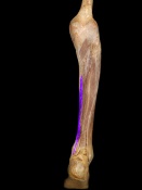
Posterior surface of the body of the tibia.
Insertion[edit | edit source]
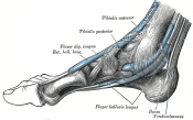
Plantar surface, base of the distal phalanges of the four lesser toes.
Nerve[edit | edit source]
Tibial nerve
Artery[edit | edit source]
Posterior tibial artery
Function[edit | edit source]
Clinical relevance[edit | edit source]
Assessment[edit | edit source]
Palpation[edit | edit source]
Power[edit | edit source]
Length[edit | edit source]
Treatment[edit | edit source]
Strengthening[edit | edit source]
Stretching[edit | edit source]
Manual techniques[edit | edit source]
Resources[edit | edit source]

|

|
File:FDL4.JPG | 
|


