Cervical Plexus: Difference between revisions
Evan Thomas (talk | contribs) No edit summary |
Kim Jackson (talk | contribs) m (Text replacement - "Category:Anatomy - Cervical Spine" to "") |
||
| (17 intermediate revisions by 5 users not shown) | |||
| Line 6: | Line 6: | ||
== Description == | == Description == | ||
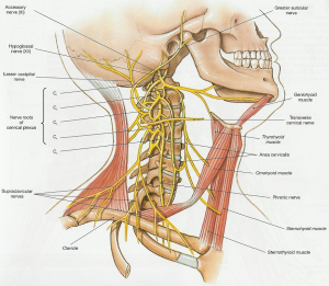
The cervical plexus is formed by the communication of the anterior divisions of the upper four cervical nerves (C1-4).<ref name="AE">http://www.anatomyexpert.com/structure_detail/6560/</ref> | The cervical plexus is formed by the communication of the anterior divisions of the upper four cervical nerves (C1-4). <ref name="AE">http://www.anatomyexpert.com/structure_detail/6560/</ref> | ||
[[Image:Cervical plexus anatomy.png|center|300px]] | |||
== Location/Path == | == Location/Path == | ||
| Line 19: | Line 14: | ||
It lies under the sternocleidomastoid (SCM) muscle, opposite the upper four cervical vertebrae. It rests upon the levator anguli scapulae and scalenus medius muscles, and emerges from the posterior border of the SCM.<ref name="AE" /> | It lies under the sternocleidomastoid (SCM) muscle, opposite the upper four cervical vertebrae. It rests upon the levator anguli scapulae and scalenus medius muscles, and emerges from the posterior border of the SCM.<ref name="AE" /> | ||
== Branches | <br> [[Image:Cervical plexus schema.png|center|500px|OCI_right_lateral_view]] | ||
== Branches and Supplied Structures == | |||
{| class="FCK__ShowTableBorders" width="40%" cellspacing="1" cellpadding="1" border="0" align="right" | |||
|- | |||
| align="right" | | |||
| {{#ev:youtube|Oj9J9b8FIIg|250}} <ref>fatcat2983472. Cervical Plexus Drawing SO GOOD!!!. Available from: http://www.youtube.com/watch?v=Oj9J9b8FIIg [last accessed 12/02/16]</ref> | |||
|} | |||
Its branches consist of a superficial and deep set. The superficial branches are the great auricular nerve, lesser occipital nerve, transverse cervical, suprasternal, and supraclavicular nerves. The deep branches are the phrenic, communicantes cervicales, communicating, and muscular.<ref name="AE" /> | Its branches consist of a superficial and deep set. The superficial branches are the great auricular nerve, lesser occipital nerve, transverse cervical, suprasternal, and supraclavicular nerves. The deep branches are the phrenic, communicantes cervicales, communicating, and muscular.<ref name="AE" /> | ||
| Line 32: | Line 35: | ||
**''Sensory'': Superior region behind auricle | **''Sensory'': Superior region behind auricle | ||
**''Motor'': None | **''Motor'': None | ||
{| class="FCK__ShowTableBorders" width="40%" cellspacing="1" cellpadding="1" border="0" align="right" | |||
|- | |||
| align="right" | | |||
| {{#ev:youtube|-Nz8-bnZGBI|250}} <ref>Kiara Rivera. Cervical Plexus Drawing and Spinal Segments - EASY. Available from: http://www.youtube.com/watch?v=-Nz8-bnZGBI [last accessed 12/02/16]</ref> | |||
|} | |||
*'''Great Auricular Nerve (C2-3)<ref name="Thompson" />''' | *'''Great Auricular Nerve (C2-3)<ref name="Thompson" />''' | ||
| Line 55: | Line 64: | ||
== Diagram == | == Diagram == | ||
[[Image:Cervical plexus diagram.PNG|center|600x400px|Cervical_plexus_diagram]] | [[Image:Cervical plexus diagram.PNG|center|600x400px|Cervical_plexus_diagram]]<div class="researchbox"> </div> | ||
== References == | == References == | ||
<references /> | <references /> <br> | ||
[[Category:Anatomy]] | |||
[[Category:Cervical Spine - Anatomy]] | |||
[[Category:Cervical Spine]] | |||
[[Category:Nerves]] | |||
[[Category:Musculoskeletal/Orthopaedics]] | |||
[[Category:Cervical Spine - Nerves]] | |||
Latest revision as of 13:40, 23 August 2019
Original Editor - Evan Thomas
Lead Editors - Evan Thomas, Laura Ritchie, Kim Jackson, WikiSysop and Leana Louw
Description[edit | edit source]
The cervical plexus is formed by the communication of the anterior divisions of the upper four cervical nerves (C1-4). [1]
Location/Path[edit | edit source]
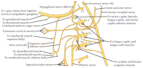
It lies under the sternocleidomastoid (SCM) muscle, opposite the upper four cervical vertebrae. It rests upon the levator anguli scapulae and scalenus medius muscles, and emerges from the posterior border of the SCM.[1]
Branches and Supplied Structures[edit | edit source]
| [2] |
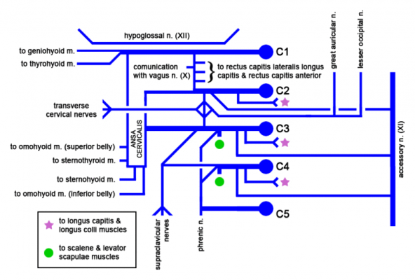
Its branches consist of a superficial and deep set. The superficial branches are the great auricular nerve, lesser occipital nerve, transverse cervical, suprasternal, and supraclavicular nerves. The deep branches are the phrenic, communicantes cervicales, communicating, and muscular.[1]
- Ansa Cervicalis (C1-3)[3]
- Superior (C1-2) & inferior (C2-3) roots form loop
- Sensory: None
- Motor: Omohyoid, sternohyoid, sternothyroid
- Lesser Occipital Nerve (C2-3)[3]
- Arises from posterior border of SCM
- Sensory: Superior region behind auricle
- Motor: None
| [4] |
- Great Auricular Nerve (C2-3)[3]
- Exits inferior to lesser occipital nerve, ascends on SCM
- Sensory: Over parotid gland and behind ear
- Motor: None
- Transverse Cervical Nerve (C2-3)[3]
- Exits inferior to greater auricular nerve, then to anterior neck
- Sensory: Anterior triangle of the neck
- Motor: None
- Supraclavicular (C2-3)[3]
- Splits into 3 branches: anterior, middle, posterior
- Sensory: Over clavicle, outer trapezius and deltoid
- Motor: None
- Phrenic Nerve (C3-5)[3]
- On anterior scalene, into thorax between subclavian artery and vein
- Sensory: Pericardium and mediastinal pleura
- Motor: Diaphragm
Diagram[edit | edit source]
References[edit | edit source]
- ↑ 1.0 1.1 1.2 http://www.anatomyexpert.com/structure_detail/6560/
- ↑ fatcat2983472. Cervical Plexus Drawing SO GOOD!!!. Available from: http://www.youtube.com/watch?v=Oj9J9b8FIIg [last accessed 12/02/16]
- ↑ 3.0 3.1 3.2 3.3 3.4 3.5 Thompson JC (2010). Netter's Concise Orthopaedic Anatomy (2nd ed). Philadelphia, PA: Saunders Elsevier.
- ↑ Kiara Rivera. Cervical Plexus Drawing and Spinal Segments - EASY. Available from: http://www.youtube.com/watch?v=-Nz8-bnZGBI [last accessed 12/02/16]


