Cartilage: Difference between revisions
No edit summary |
No edit summary |
||
| Line 74: | Line 74: | ||
Participation in certain sports appear to increase risk of [[osteoarthritis]] (result from breakdown of joint cartilage). Activities that involve torsional loading , fast acceleration and deceleration , repetitive high impact and high level of participation increase risk of osteoarthritis. Increased risks of osteoarthritis are related to excessive exercise or abnormal joint loading But some levels of loading and exercise are beneficial for joint health as exercise enhances production of matrix molecules, which can have a positive effect on joint health. Therefore exercise has both beneficial and detrimental effects on cartilage. Exercise can promote positive biomechanical changes, reduce pain and increase function of people with arthritis but it must be noted that sports injuries can be a contributing factor to developing osteoarthritis .<ref name="Carol" /> | Participation in certain sports appear to increase risk of [[osteoarthritis]] (result from breakdown of joint cartilage). Activities that involve torsional loading , fast acceleration and deceleration , repetitive high impact and high level of participation increase risk of osteoarthritis. Increased risks of osteoarthritis are related to excessive exercise or abnormal joint loading But some levels of loading and exercise are beneficial for joint health as exercise enhances production of matrix molecules, which can have a positive effect on joint health. Therefore exercise has both beneficial and detrimental effects on cartilage. Exercise can promote positive biomechanical changes, reduce pain and increase function of people with arthritis but it must be noted that sports injuries can be a contributing factor to developing osteoarthritis .<ref name="Carol" /> | ||
= Anatomy, Cartilage = | |||
Chang IR, Martin A. | |||
Publication Details | |||
== Introduction == | |||
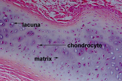
Cartilage has many functions including the ability to resist compressive forces, enhance bone resilience, and provide support on bony areas where there is a need for flexibility. The primary cell that makes cartilage is the chondrocyte which resides within the lacunae. The matrix of cartilage consists of fibrous tissue and various combinations of proteoglycans and glycosaminoglycans. Cartilage once synthesized, lacks lymphatic or blood supply and the movement of waste and nutrition is chiefly via diffusion to and from adjacent tissues. Cartilage, like bone, is surrounded by a perichondrium-like fibrous membrane. This layer is not efficient at regeneration of cartilage. Hence, its recovery is slow after injury. The lack of active blood flow is the major reason any injury to cartilage takes a long time to heal. Cartilage has no nerve innervation, and hence there is no sensation when it is injured or damaged. When there is calcification of cartilage, the chondrocytes die. This is followed by replacement of cartilage with bone-like tissue. Unlike bone, cartilage does not have calcium in the matrix. Instead, it contains high amounts of chondroitin, which is the material that provides elasticity and flexibility. | |||
== Structure and Function == | |||
There are several types of cartilage found in the human body, and their structure and relevant function depend on this variation. | |||
'''Hyaline Cartilage''' | |||
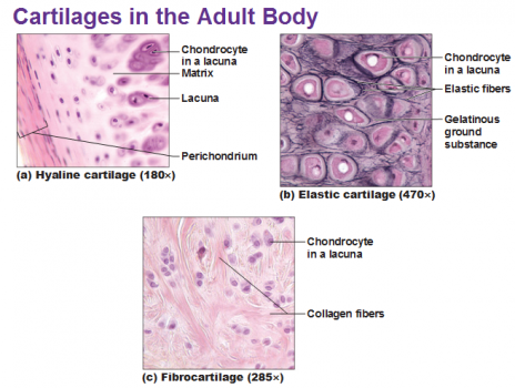
Hyaline cartilage is the most copious type of cartilage in the human body.[1] It has a pale blue-white color and is smooth to the touch. It is primarily composed of type II collagen and proteoglycans. The surface is usually moist, but with age, the cartilage becomes dry, thinner, and more yellow. Hyaline cartilage is usually found in the trachea, nose, epiphyseal growth plate, sternum, and ventral segments of the ribs. Hyaline cartilage produces a resilient surface with minimal friction. It also has an excellent ability to resist compressive forces at sites of bone articulation.[2] | |||
'''Elastic Cartilage''' | |||
This cartilage appears a dull yellow and is most commonly found in the larynx, ear, epiglottis, and eustachian tube. It is also surrounded by a perichondrium-like layer. It provides flexibility and is resilient to pressure.[3] | |||
'''Fibrocartilage''' | |||
This is abundant in type 1 collagen and contains significantly less proteoglycan than hyaline cartilage. It can resist high degrees of tension and compression. It is commonly found in tendons, ligaments, intervertebral discs, articular surfaces of some bones, and in menisci. Unlike other cartilage, it has no perichondrium.[4] | |||
== Embryology == | |||
Cartilage is formed from the mesoderm germ layer by the process known as chondrogenesis.[5] Mesenchyme differentiates into chondroblasts which are the cells that secrete the major components of the extracellular matrix. The most important of these components for cartilage formation being aggrecan and type II collagen. Once initial chondrification occurs, the immature cartilage grows mainly by developing into a more mature state since it cannot grow by mitosis. There is minimal cell division in cartilage; therefore, the size and mass of cartilage do not change significantly after initially chondrification. | |||
== Blood Supply and Lymphatics == | |||
Cartilage is avascular. This characteristic of cartilage is paramount during the discussion and management of diseases affecting cartilage. Since there is no direct blood supply, chondrocytes receive nourishment via diffusion from the surrounding environment. The compressive forces that regularly act on cartilage also increase the diffusion of nutrients. This indirect process of receiving nutrients is a major factor in the slow turnover of the extracellular matrix and lack of repair seen in cartilage. | |||
== Nerves == | |||
Cartilage does not contain nerves; it is aneural.[6] The pain, if any, associated with pathology involving cartilage is most commonly due to irritation of surrounding structures, for example, inflammation of the joint and bone in osteoarthritis. | |||
== Muscles == | |||
Fibrocartilage is a major component of entheses, which is the connective tissue between muscle tendon or ligament and bone. The fibrocartilaginous enthesis consists of 4 transition zones as it progresses from tendon to bone.[7] These transition zones are listed in order of progression from muscle to bone. | |||
# Longitudinal fibroblasts and a parallel arrangement of collagen fibers are found at the tendinous area | |||
# A fibrocartilaginous region where the main type of cells present transitions from fibroblast to chondrocytes | |||
# A region called the "blue line" or "tide mark" due to an abrupt transition from cartilaginous to calcified fibrocartilage | |||
# Bone | |||
== Physiologic Variants == | |||
Literature shows many anatomical variants of cartilage, and in many cases, this can affect the pathology associated. For example, a study showed a significant correlation between novel genetic variants in cartilage thickness and the incidence of hip osteoarthritis.[8] | |||
== Clinical Significance == | |||
An immense variety of clinical pathology exists involving cartilage, for example, [[osteoarthritis]], spinal [[Disc Herniation|disc herniation]], traumatic rupture/detachment, achondroplasia, costochondritis, neoplasm, and many others. These result from a variety of degenerative, inflammatory, and congenital causes. | |||
== Current thinking on articular repair and regeneration == | == Current thinking on articular repair and regeneration == | ||
Revision as of 13:02, 10 February 2020
Top Contributors - Esraa Mohamed Abdullzaher, Lucinda hampton, Kim Jackson, George Prudden, Joao Costa and Sai Kripa
Introduction[edit | edit source]
Cartilage is a non-vascular type of supporting connective tissue that is found throughout the body .
- Cartilage is a flexible connective tissue that differs from bone in several ways; it is avascular and its microarchitecture is less organized than bone.
- Cartilage is not innervated and therefore relies on diffusion to obtain nutrients. This causes it to heal very slowly.
- The main cell types in cartilage are chondrocytes, the ground substance is chondroitin sulfate, and the fibrous sheath is called perichondrium.
- There are three types of cartilage: hyaline, fibrous, and elastic cartilage.
- Hyaline cartilage is the most widespread type and resembles glass. In the embryo, bone begins as hyaline cartilage and later ossifies.
- Fibrous cartilage has many collagen fibers and is found in the intervertebral discs and pubic symphysis.
- Elastic cartilage is springy, yellow, and elastic and is found in the internal support of the external ear and in the epiglottis[1]
Cartilage Structure [edit | edit source]
Cartilage is a dense structure, that resembles a firm gel, made up of collagen and elastic fibres. It contains polysacchride derivaites called chondroitin sulfates which complex with protein in the ground substance forming proteoglycan. The matrix is produced by cells call chrondroblasts which form chrondocytes and can be found in small chambers called lacuna
Cartilage is separated from the surrounding tissues by perichondrium which consist of two layers:
- Outer Fibrous Layer : Which provide protection , mechanical support and attaches the cartilage to other structures.
- Inner Cellular : It Is Important in the growth and maintenance of cartilage .[3]
Types of Cartilage[edit | edit source]
There are three types of cartilage and they all have slightly different structures and function
Hyaline Cartilage[edit | edit source]
Hyaline Cartilage has a smooth surface and is the most common of the three types of cartilage. It has a matrix that contains closely packed collagen fibers, making it tough but slightly flexible. It consists of a bluish-white, shiny ground elastic material, whose matrix contains chondtoitin sulphate, with many fine collagen fibrils and chrondrocytes. Because of its smooth surfaces it allows tissues to slide/glide more easily, as well as providing flexibility and support
Example : Connection between ribs and sternum, nasal cartilage and articular cartilage (which covers opposing bone surfaces in many joints).
Fibrocartilage[edit | edit source]
Fibrocartilage is the toughest of the three types of cartilage. It has no perichondrium and has a matrix that contains dense bundles of collagen fibers embedded with chrondrocytes, making it durable and tough. This makes it perfect to provide support and rigidity
Example : Intervertebral discs (between spinal vertebrae), Menisci (cartilage pads of the knee joint), the callus (formed at the ends of bones at the site of a fracture), between the Pubic Symphysis and at the junction where tendons insert into bone.
Elastic Cartilage[edit | edit source]
Elastic cartilage provides support. It has a yellowish colour and is surrounded by a perichnodrium. Chrondrocytes are located between a network of threadlike elastic fibres, the abundance of elastic fibres makes it flexible and resilient.
Example : the auricle of the outer ear .
Cartilage Growth [edit | edit source]
The growth of cartilage is a slow process and occurs by the division of cells. Both intersitial and appositional growth occur during development but neither of them occur in normal adults but under unusual circumstances appositional growth may occur as in cartilage damage (minor damage) .[3]
Interstitial Growth[edit | edit source]
Chondrocytes responsible for cell division and daughter cells produce the matrix. This occurs during embryonic level and continues until adolescence.
Appostional Growth[edit | edit source]
Cells of inner layer of perichondrium undergo division and inner most cells differentiate into chondroblasts which produce the matrix, they then mature into mature chondrocytes. The main difference between chondrocytes and chondroblasts is that chondroblasts secrete the extracellular matrix of the cartilage whereas chondrocytes are involved in the maintenance of the cartilage.[4]
Remodelling of Cartilage[edit | edit source]
This occurs predominantly by changes and the rearrangement of the collagen matrix in response to load.
Mechanical Behaviour of Articular Cartilage[edit | edit source]
The mechanical behaviour depend on interaction of its component : proteoglycan, collagen and interstitial fluid. In an aqueous environment , proteogylcans are polyanionic which means the molecule has negatively charged sites that arise from sulfate and carboxyl. In solution, the mutual repulsion of these negative charges causes the aggregated proteogylcan to spread out and occupy a large volume .
In the cartilage matrix, the volume occupied by proteogylcan aggregates is limited by the network of collagen fibres. when the cartilage is commpressed the negatively charged sites are pushed together increasing the mutual repulsion force adding to the compressive stiffness of the cartilage. During this process non-aggregated protoegylcans are not affected by the compressive load since they are not easily trapped in the cartilage matrix .Damage to the collagen framework reduces compressive stiffness .
The mechanical response of the cartilage is strongly tied to the application of pressure differences and the flow of fluid through the tissue as, when deformed, the fluid flows across the cartilage and articular surface .
Biphasic model of cartilage[edit | edit source]
All of the solid components of the cartilage (lipid, proteogylcans,cells and collagen ) are grouped together to form the solid component of the matrix and the interstitial fluid, that moves freely, forms the fluid component.
Exercise and Cartilage Health[edit | edit source]
Participation in certain sports appear to increase risk of osteoarthritis (result from breakdown of joint cartilage). Activities that involve torsional loading , fast acceleration and deceleration , repetitive high impact and high level of participation increase risk of osteoarthritis. Increased risks of osteoarthritis are related to excessive exercise or abnormal joint loading But some levels of loading and exercise are beneficial for joint health as exercise enhances production of matrix molecules, which can have a positive effect on joint health. Therefore exercise has both beneficial and detrimental effects on cartilage. Exercise can promote positive biomechanical changes, reduce pain and increase function of people with arthritis but it must be noted that sports injuries can be a contributing factor to developing osteoarthritis .[6]
Anatomy, Cartilage[edit | edit source]
Chang IR, Martin A.
Publication Details
Introduction[edit | edit source]
Cartilage has many functions including the ability to resist compressive forces, enhance bone resilience, and provide support on bony areas where there is a need for flexibility. The primary cell that makes cartilage is the chondrocyte which resides within the lacunae. The matrix of cartilage consists of fibrous tissue and various combinations of proteoglycans and glycosaminoglycans. Cartilage once synthesized, lacks lymphatic or blood supply and the movement of waste and nutrition is chiefly via diffusion to and from adjacent tissues. Cartilage, like bone, is surrounded by a perichondrium-like fibrous membrane. This layer is not efficient at regeneration of cartilage. Hence, its recovery is slow after injury. The lack of active blood flow is the major reason any injury to cartilage takes a long time to heal. Cartilage has no nerve innervation, and hence there is no sensation when it is injured or damaged. When there is calcification of cartilage, the chondrocytes die. This is followed by replacement of cartilage with bone-like tissue. Unlike bone, cartilage does not have calcium in the matrix. Instead, it contains high amounts of chondroitin, which is the material that provides elasticity and flexibility.
Structure and Function[edit | edit source]
There are several types of cartilage found in the human body, and their structure and relevant function depend on this variation.
Hyaline Cartilage
Hyaline cartilage is the most copious type of cartilage in the human body.[1] It has a pale blue-white color and is smooth to the touch. It is primarily composed of type II collagen and proteoglycans. The surface is usually moist, but with age, the cartilage becomes dry, thinner, and more yellow. Hyaline cartilage is usually found in the trachea, nose, epiphyseal growth plate, sternum, and ventral segments of the ribs. Hyaline cartilage produces a resilient surface with minimal friction. It also has an excellent ability to resist compressive forces at sites of bone articulation.[2]
Elastic Cartilage
This cartilage appears a dull yellow and is most commonly found in the larynx, ear, epiglottis, and eustachian tube. It is also surrounded by a perichondrium-like layer. It provides flexibility and is resilient to pressure.[3]
Fibrocartilage
This is abundant in type 1 collagen and contains significantly less proteoglycan than hyaline cartilage. It can resist high degrees of tension and compression. It is commonly found in tendons, ligaments, intervertebral discs, articular surfaces of some bones, and in menisci. Unlike other cartilage, it has no perichondrium.[4]
Embryology[edit | edit source]
Cartilage is formed from the mesoderm germ layer by the process known as chondrogenesis.[5] Mesenchyme differentiates into chondroblasts which are the cells that secrete the major components of the extracellular matrix. The most important of these components for cartilage formation being aggrecan and type II collagen. Once initial chondrification occurs, the immature cartilage grows mainly by developing into a more mature state since it cannot grow by mitosis. There is minimal cell division in cartilage; therefore, the size and mass of cartilage do not change significantly after initially chondrification.
Blood Supply and Lymphatics[edit | edit source]
Cartilage is avascular. This characteristic of cartilage is paramount during the discussion and management of diseases affecting cartilage. Since there is no direct blood supply, chondrocytes receive nourishment via diffusion from the surrounding environment. The compressive forces that regularly act on cartilage also increase the diffusion of nutrients. This indirect process of receiving nutrients is a major factor in the slow turnover of the extracellular matrix and lack of repair seen in cartilage.
Nerves[edit | edit source]
Cartilage does not contain nerves; it is aneural.[6] The pain, if any, associated with pathology involving cartilage is most commonly due to irritation of surrounding structures, for example, inflammation of the joint and bone in osteoarthritis.
Muscles[edit | edit source]
Fibrocartilage is a major component of entheses, which is the connective tissue between muscle tendon or ligament and bone. The fibrocartilaginous enthesis consists of 4 transition zones as it progresses from tendon to bone.[7] These transition zones are listed in order of progression from muscle to bone.
- Longitudinal fibroblasts and a parallel arrangement of collagen fibers are found at the tendinous area
- A fibrocartilaginous region where the main type of cells present transitions from fibroblast to chondrocytes
- A region called the "blue line" or "tide mark" due to an abrupt transition from cartilaginous to calcified fibrocartilage
- Bone
Physiologic Variants[edit | edit source]
Literature shows many anatomical variants of cartilage, and in many cases, this can affect the pathology associated. For example, a study showed a significant correlation between novel genetic variants in cartilage thickness and the incidence of hip osteoarthritis.[8]
Clinical Significance[edit | edit source]
An immense variety of clinical pathology exists involving cartilage, for example, osteoarthritis, spinal disc herniation, traumatic rupture/detachment, achondroplasia, costochondritis, neoplasm, and many others. These result from a variety of degenerative, inflammatory, and congenital causes.
Current thinking on articular repair and regeneration[edit | edit source]
A biological approach to cartilage damage is challenging due to its' inherent limited healing potential. Various options have been made available over the years trying to address these issues. New technique have merits and demerits. Stem cells therapy is a strong promise in the treatment of cartilage defects and osteoarthritis.
Stem cells(SC), in particular mesenchymal SCs, are expected to revolutionise the treatment for cartilage defects and osteoarthritis in the near future. It is hoped that the whole cartilage can be repaired not just focal defects.[7]
The below video delves into the regeneration aspirations
References[edit | edit source]
- ↑ Lumen learning Cartilage Available from:https://courses.lumenlearning.com/boundless-ap/chapter/cartilage/ (last accessed 10.2.2020)
- ↑ https://www.youtube.com/watch?v=mr5JI8Q8dc8
- ↑ 3.0 3.1 3.2 Fredric H.Martini ,Judi Nath ,Edwen Bartholomew , Charles M.Seiger ,Damian Hill . Fundamentals of Anatomy and Physiology .9th ed , 2011 .
- ↑ Eoediaa Differences between chondroblasts and chondrocytes Available from: https://pediaa.com/difference-between-chondroblasts-and-chondrocytes/ (last accessed 31.5.2019)
- ↑ https://www.youtube.com/watch?v=tS5S8BoVN-4
- ↑ 6.0 6.1 Carol A.Oatis . kinesiology the mechanics and pathomechanics of human movement , 2003 .
- ↑ Karuppal R. Current concepts in the articular cartilage repair and regeneration. J Orthop. 2017;14(2):A1–A3. Published 2017 May 19. doi:10.1016/j.jor.2017.05.001 Available from:https://www.ncbi.nlm.nih.gov/pmc/articles/PMC5440635/ (last accessed 31.5.2019)
- ↑ Sportology Cartilage repair basics Available from: https://www.youtube.com/watch?v=CxhFhidWn6w (last accessed 31.5.2019)


