Calcaneal Spurs: Difference between revisions
No edit summary |
No edit summary |
||
| Line 82: | Line 82: | ||
== Recent Related Research (from [http://www.ncbi.nlm.nih.gov/pubmed/ Pubmed]) == | == Recent Related Research (from [http://www.ncbi.nlm.nih.gov/pubmed/ Pubmed]) == | ||
<br> | |||
<div class="researchbox"> | <div class="researchbox"><rss>http://www.ncbi.nlm.nih.gov/entrez/eutils/erss.cgi?rss_guid=1NIs-Zi-UW0qwJBfJz_3MSjudXvn9v2gqwmkKwdQHFZeSS0kYp|charset=UTF-8|short|max=10</rss></div> | ||
<rss> | |||
</div> | |||
== References == | == References == | ||
Revision as of 10:24, 29 June 2014
Original Editors - Caro De Koninck
Top Contributors - Ivakhnov Sergei, Caro De Koninck, Sheik Abdul Khadir, Scott Cornish, Admin, Simisola Ajeyalemi, Kim Jackson, Rachael Lowe, Joao Costa, Wanda van Niekerk, 127.0.0.1, Evan Thomas, Naomi O'Reilly and Priyanka Chugh
Definition/Description[edit | edit source]
A calcaneal spur occurs when there is a bone spur, a small bony outgrowth, formed on the heel bone. A more common name for calcaneal spur is heel spur. Calcaneal spurs can be located at the back of the heel or under the sole. Spurs at the back are often associated with Achilles tendonitis, while spurs under the sole are associated with plantar fasciitis.

Epidemiology /Etiology[edit | edit source]
The etiology of the spur have been debated. At the beginning of the twentieth century, gonorrhea was considered a prime ethiological factor. Heredity, metabolic disorders, tuberculosis, systemic inflammatory diseases and many other disorders have also been implicated. Now abnormal biomechanics (excessive pronation) enjoys wide support as the prime etiological factor for the painful plantar heel and the inferior calcaneal spur. The spur is thought to be a result of the biomechanical fault and an incidental finding when associated with the painful plantar heel. [1]
Characteristics/Clinical Presentation[edit | edit source]
The painful heel is a relatively common foot problem but calcaneal spurs are not considered a primary cause of heel pain. Most heel pain patients are middle-aged adults. Many of them are obese, so obesity can be considered a risk factor.
Not all heel spurs cause symptoms but when they to people often experience more pain during weight-bearing activities, in the morning or after a period of rest. The pain however is not the result of pressure of weight on the top of the spur but the experienced pain is coming from an inflammation around tendons where they attach to the bone.
Differential Diagnosis [2][edit | edit source]
- Calcaneal stress fracture
- Nerve entrapment
- Peroneal Tendinopathy
- Syndrome- sinus tarsi / Tarsal tunnel
- Sever disease (Calcaneal apophysitis)
- Haglund Deformity (With or without busitis)
Diagnostic Procedures[edit | edit source]
The diagnose is based on the patient's history and on results of the physical examination. The suspicion diagnose is confirmed mostly in the X-ray on the calcaneus. Rarely MRI may be required.
Clinical Examination
[edit | edit source]
- Posture of foot (with and without weight bearing & with and without footwear)
- Tenderness (Site/ Severity)
- Range of Motion in ankle and foot
- Muscle Strength
- Sensation
- Gait
Medical Management
[edit | edit source]
- Non steroidal anti inflammatory drugs
- Steroids - oral /injections
Non Medical Management[edit | edit source]
Physiotherapy[edit | edit source]
Calcaneal spurs, both upper and lower spurs, are treated with conventional physiotherapy.
- Conventional therapy includes ultrasound, Laser, passive and active stretching and strengthening of the muscles of the legs and cold and warmth applications (Contrast Bath). The aim is to eliminate the inflammation surrounding the spur. This kind of treatment may take 6 to 12 months.
- Another proven therapy (D'andrea greve et al., 2009, B) [3] is radial shockwave therapy. This method consists of very high-energy mechanical waves, pointed at the calcaneal spur. By the growth of blood vessels the inflammation shall be taken away.
- D'andrea greve et al.(2009, B) [3] claim that their is no difference between both methods of treatment.
However there are different studies who claim ESWT is not effective in the treatment of plantar fasciitis. (Buchanan et al. 2002, A2; Haake et al. 2003, A2) [4] [5] Because of this discrepancy between studies, further support for an effective treatment with ESWT is needed. According to De Vera Barredo et al.(2007, A1) [6] night splints, massage, taping, acupuncture, walking casts, laser therapy and cryotherapy are more means to help the patient.
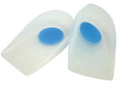
Orthosis[edit | edit source]
- Night Splints
- Heel inserts
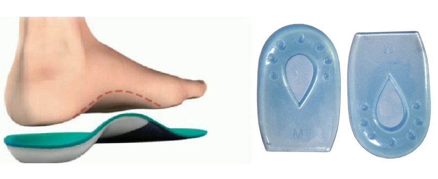
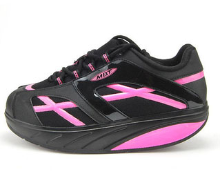
- Foot wear Modification


When neither of these treatments take away the problem, more aggressive techniques like surgery can be employed.
Resources
[edit | edit source]
Buzzle.com, Intelligent Life on the web. Calcaneal spur. http://www.buzzle.com/articles/calcaneal-spur.html (accessed 15 november 2010)
MedicineNet.com. Definition of calcaneal spur. http://www.medterms.com/script/main/art.asp?articlekey=7095 (accessed 15 november 2010)
Recent Related Research (from Pubmed)[edit | edit source]
References[edit | edit source]
- ↑ Edmund M, Kosmahl PT, MS, Herbert E, Kosmahl DPM. Painful Plantar Heel, PlantarfckLRFasciitis, and Calcaneal Spur: Etiology and treatment. J Orthop Sports Phys Ther 1987; 9(1)
- ↑ PRISCILLA TU, DO, and JEFFREY R. BYTOMSKI, DO, Duke University, Durham, North Carolina:Diagnosis of Heel Pain:from the American Family Physician Web site at www.aafp.org/afp.
- ↑ 3.0 3.1 D'andrea Greve JM, Grecco MV, Santos-Silva PR. Comparison of radial shockwaves andfckLRconventional physiotherapy for treating plantar fasciitis. Clinics 2009; 64(2)fckLRhttp://www.scielo.br/scielo.php?script=sci_arttext&amp;amp;amp;amp;amp;amp;amp;amp;amp;pid=S1807-fckLR59322009000200006&amp;amp;amp;amp;amp;amp;amp;amp;amp;lng=en&amp;amp;amp;amp;amp;amp;amp;amp;amp;nrm=iso&amp;amp;amp;amp;amp;amp;amp;amp;amp;tlng=en (accessed 20 november 2010)
- ↑ Buchanan J, Buchbinder R, Forbes A, Gordon J, Prabaharan V, Ptatsnik R. UltrasoundguidedfckLRextracorporeal shockwave therapy for plantar fasciitis. JAMA 2002; 288: 1364-1372fckLRhttp://jama.ama-assn.org/content/288/11/1364.full.pdf+html (accessed 27 december 2010)
- ↑ Buch M, Haake M, Schoellner C, Goebel F, Vogel M, Mueller I, Hausdorf J, Zamzow K,fckLRSchade-Brittinger C, Mueller H-H. Extracorporeal shock wave therapy for plantar fasciitis:fckLRrandomised controlled multicentre trial. BMJ 2003; 327fckLRhttp://www.bmj.com/content/327/7406/75.full.pdf (accessed 27 december 2010)
- ↑ De Vera Barredo R, Menna D, Farris JW. An evaluation of research evidence for selectedfckLRphysical therapy interventions for plantar fasciitis. J Phy The Sci 2007; 19: 41-56fckLRhttp://www.jstage.jst.go.jp/article/jpts/19/1/41/_pdf (accessed 20 november 2010)

