Calcaneal Spurs: Difference between revisions
No edit summary |
No edit summary |
||
| Line 14: | Line 14: | ||
== Clinically Relevant Anatomy == | == Clinically Relevant Anatomy == | ||
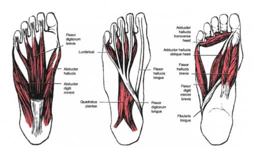
[[Image:Intrinsic foot muscles.png|border|right|Intrinsic foot muscles]]<br> | [[Image:Intrinsic foot muscles.png|border|right|600x300px|Intrinsic foot muscles]]<br> | ||
M. Soleus<br>M. Gastrocnemius<br>M. Plantaris <br>M. Abductor Digiti minimi<br>M. Flexor digitorum brevis<br>M. Extensor digitorum brevis<br>M. Abductor hallucis<br>M. Extensor hallucis brevis<br>M. Quadratus plantae<br>Plantar fascia<br><br> | M. Soleus<br>M. Gastrocnemius<br>M. Plantaris <br>M. Abductor Digiti minimi<br>M. Flexor digitorum brevis<br>M. Extensor digitorum brevis<br>M. Abductor hallucis<br>M. Extensor hallucis brevis<br>M. Quadratus plantae<br>Plantar fascia<br><br> | ||
All of these structures are in a position to exert a traction force on the tuberosity and adjacent regions of the calcaneus, especially when excessive or abnormal pronation occurs. The origin of the spurs appears to be repetitive trauma that produced microtears in the plantar fascia near its attachment and the attempted repair led to inflammation which is responsible for the release and the maintenance of the symptoms.<ref>Gill LH. Plantar fasciitis: diagnosis and conservative management. J Am Acad Orthop Surg, 1997</ref>,<ref>McCarthy DJ, Gorecki GE: The anatomical basis of inferior calcaneal lesions. J Am Podiatry Assoc 69527-536,1979</ref>,<ref>Young CC, Rutherford DS, Niedfeldt MW. Treatment of plantar fasciitis. Am | All of these structures are in a position to exert a traction force on the tuberosity and adjacent regions of the calcaneus, especially when excessive or abnormal pronation occurs. The origin of the spurs appears to be repetitive trauma that produced microtears in the plantar fascia near its attachment and the attempted repair led to inflammation which is responsible for the release and the maintenance of the symptoms.<ref>Gill LH. Plantar fasciitis: diagnosis and conservative management. J Am Acad Orthop Surg, 1997</ref>,<ref>McCarthy DJ, Gorecki GE: The anatomical basis of inferior calcaneal lesions. J Am Podiatry Assoc 69527-536,1979 (level of evidence: 2C)</ref>,<ref>Young CC, Rutherford DS, Niedfeldt MW. Treatment of plantar fasciitis. Am | ||
Fam Physician 2001</ref>,<ref>Heyd, Reinhard, et al. "Radiation therapy for painful heel spurs." Strahlentherapie und Onkologie 183.1 (2007): 3-9</ref> | Fam Physician 2001 (level of evidence: 5)</ref>,<ref>Heyd, Reinhard, et al. "Radiation therapy for painful heel spurs." Strahlentherapie und Onkologie 183.1 (2007): 3-9. (level of evidence: 1B)</ref> | ||
== Epidemiology /Etiology == | == Epidemiology /Etiology == | ||
Revision as of 11:07, 19 May 2016
Original Editors
Top Contributors - Ivakhnov Sergei, Caro De Koninck, Sheik Abdul Khadir, Scott Cornish, Admin, Simisola Ajeyalemi, Kim Jackson, Rachael Lowe, Joao Costa, Priyanka Chugh, Wanda van Niekerk, 127.0.0.1, Evan Thomas and Naomi O'Reilly
Search Strategy[edit | edit source]
add text here related to databases searched, keywords, and search timeline
Definition/Description[edit | edit source]
A calcaneal spur, or commonly known as a heel spur, occurs when there is a bone spur (a bony outgrowth) formed on the heel bone. Calcaneal spurs can be located at the back of the heel (dorsal heel spur) or under the sole (plantar heel spur). The dorsal spurs are often associated with achilles Tendinopathy, while spurs under the sole are associated with Plantar fasciitis.
The apex of the spur lies either within the origin of the planter fascia (on the medial tubercle of the calcaneus) or superior to it (in the origin of the flexor digitorum brevis muscle). The relationship between spur formation, the medial tubercle of the calcaneus and intrinsic heel musculature results in a constant pulling effect on the plantar fascia consequent a inflammatory process[1].
Clinically Relevant Anatomy[edit | edit source]
M. Soleus
M. Gastrocnemius
M. Plantaris
M. Abductor Digiti minimi
M. Flexor digitorum brevis
M. Extensor digitorum brevis
M. Abductor hallucis
M. Extensor hallucis brevis
M. Quadratus plantae
Plantar fascia
All of these structures are in a position to exert a traction force on the tuberosity and adjacent regions of the calcaneus, especially when excessive or abnormal pronation occurs. The origin of the spurs appears to be repetitive trauma that produced microtears in the plantar fascia near its attachment and the attempted repair led to inflammation which is responsible for the release and the maintenance of the symptoms.[2],[3],[4],[5]
Epidemiology /Etiology[edit | edit source]
The etiology of the spur has been debated. At the beginning of the twentieth century, gonorrhea was considered a prime ethiological factor. Heredity, metabolic disorders, tuberculosis, systemic inflammatory diseases and many other disorders have also been implicated. Now abnormal biomechanics (excessive or abnormal pronation) enjoys wide support as the prime etiological factor for the painful plantar heel and the inferior calcaneal spur. The spur is thought to be a result of the biomechanical fault and an incidental finding when associated with the painful plantar heel.
The most common etiology involves abnormal pronation with resultant increased tension forces developed in the structures attaching in the region of the calcaneal tuberosity.
Asymptomatic heel spurs are relatively common in the normal, adult population. One epidemiologic study found that 11% of the adult U.S. population demonstrated a calcaneal spur as an incidental radiographic finding.[6]
Characteristics/Clinical Presentation[edit | edit source]
The painful heel is a relatively common foot problem but calcaneal spurs are not considered a primary cause of heel pain. A calcaneal spur is caused by long-term stress on the plantar fascia and muscles on the foot and may develop as a reaction to plantar fasciitis.[7]
The pain, mostly localised in the area of the medial process of the calcaneal tuber, is caused by pressure in the region of the plantar aponeurosis attachment to the calcaneal bone. The condition may exist without producing symptoms, or it may become very painful, even disabling.[8]
Most heel pain patients are middle-aged adults. Some of them are obese, so obesity can be considered a risk factor. Not all heel spurs cause symptoms and are often painless, but when they do cause symptoms people often experience more pain during weight-bearing activities, in the morning or after a period of rest. The experienced pain, however, is not the result of pressure of weight on the top of the spur but comes from an inflammation around tendons where they attach to the bone.
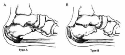
Two types of calcaneal spurs may be distinguished. Type A spurs are superior to the plantar fascia insertion, and type B spurs stretch forward from the plantar fascia insertion to extend distally within the plantar fascia.
The mean spur length for type A calcaneal spurs is significantly longer statistically than the mean spur length for type B spurs, while patients with type B spurs reported more severe clinical pain.[9]
Differently, spurs can be classified into three types:
1. There are those which are large in size but are asymptomatic, because the angle of growth is such that the spur does not become a weight-bearing point and/or the inflammatory changes have been arrested.[10]
2. Secondly, there are those which are large as well as painful upon weight-bearing, because the pitch of the calcaneus has been changed by a depression of the longitudinal arch and, as a result, the spur may become a weight-bearing point, sometimes causing intractable refractory pain.[11]
3. Lastly, there are those which have only a rudiment of proliferation and whose outline is irregular and jagged, usually accompanied by an area of decreased density around the origin of the plantar fascia, indicating a subacute inflammatory process. All calcaneal spurs undoubtedly begin in this manner, but only a few become symptomatic at this stage, because the etiologic factors are acute.[12]
Differential Diagnosis[edit | edit source]
add text here
Diagnostic Procedures[edit | edit source]
add text here related to medical diagnostic procedures
Outcome Measures[edit | edit source]
add links to outcome measures here (also see Outcome Measures Database)
Examination[edit | edit source]
add text here related to physical examination and assessment
Medical Management
[edit | edit source]
add text here
Physical Therapy Management
[edit | edit source]
add text here
Key Research[edit | edit source]
add links and reviews of high quality evidence here (case studies should be added on new pages using the case study template)
Resources
[edit | edit source]
add appropriate resources here
Clinical Bottom Line[edit | edit source]
add text here
Recent Related Research (from Pubmed)[edit | edit source]
see tutorial on Adding PubMed Feed
Extension:RSS -- Error: Not a valid URL: Feed goes here!!|charset=UTF-8|short|max=10
References[edit | edit source]
see adding references tutorial.
- ↑ Johal KS .,‘Plantar fasciitis and the calcaneal spur: Fact or fiction?’., Foot Ankle Surg.,18 March 2012
- ↑ Gill LH. Plantar fasciitis: diagnosis and conservative management. J Am Acad Orthop Surg, 1997
- ↑ McCarthy DJ, Gorecki GE: The anatomical basis of inferior calcaneal lesions. J Am Podiatry Assoc 69527-536,1979 (level of evidence: 2C)
- ↑ Young CC, Rutherford DS, Niedfeldt MW. Treatment of plantar fasciitis. Am Fam Physician 2001 (level of evidence: 5)
- ↑ Heyd, Reinhard, et al. "Radiation therapy for painful heel spurs." Strahlentherapie und Onkologie 183.1 (2007): 3-9. (level of evidence: 1B)
- ↑ McCarthy DJ, Gorecki GE: The anatomical basis of inferior calcaneal lesions. J Am Podiatry Assoc 69527-536,1979 (level of evidence: 2C)
- ↑ E.K. Agyekum., “Heel pain: A systematic review”., Chinese Journal of Traumatology., 2015 (level of evidence 1A)
- ↑ B. Jasiak-Tyrkalska., ‘Efficacy of two different physiotherapeutic preocedures in comprehensive therapy of plantar calcaneal spur’., Fizjoterapia Polska., January 2007 (level of evidence: 1B)
- ↑ Zhou, Binghua, et al. "Classification of Calcaneal Spurs and Their Relationship With Plantar Fasciitis." The Journal of Foot and Ankle Surgery 54.4 (2015): 594-600. (level of evidence: 3A)
- ↑ Henri L. Duvries., “Heel Spur (Calcaneal Spur)”., AMA Arch Surg., (level of evidence: 3A)
- ↑ Henri L. Duvries., “Heel Spur (Calcaneal Spur)”., AMA Arch Surg., (level of evidence: 3A)
- ↑ Henri L. Duvries., “Heel Spur (Calcaneal Spur)”., AMA Arch Surg., (level of evidence: 3A)


