Biceps Femoris
Original Editor - Evan Thomas
Top Contributors - Sai Kripa, Evan Thomas, Kim Jackson, Joao Costa, Ahmed M Diab, 127.0.0.1, George Prudden and WikiSysop
Description[edit | edit source]
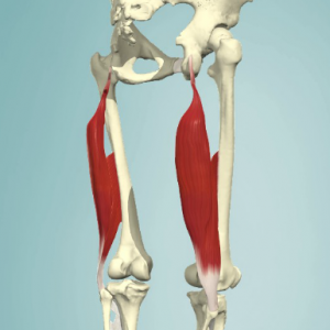
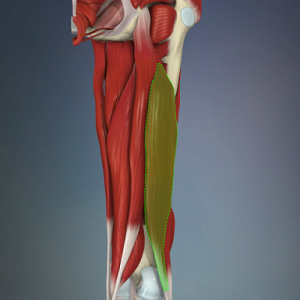
Biceps femoris is a muscle of the posterior compartment of the thigh, and lies in the posterolateral aspect. It arises proximally by two 'heads', termed the 'long head' (superficial) and the 'short head' (deep). It is part of the hamstrings.[1]
| [2] | [1] |
|---|
Anatomy[edit | edit source]
Origin[edit | edit source]
- Long head: ischial tuberosity[3]
- Short head: linea aspera and lateral supracondylar line of the femur[3]
Insertion[edit | edit source]
- Lateral aspect of fibular head[3]
Nerve Supply[edit | edit source]
- Long head: tibial division of the sciatic nerve (L5-S2)[3]
- Short head: common peroneal division of the sciatic nerve[3]
Blood Supply[edit | edit source]
- Perforating branches of profunda femoris, inferior gluteal, and medial circumflex femoral arteries[3]
Function[edit | edit source]
Actions[edit | edit source]
- Long head: flexes the knee, extends hip, laterally rotates lower leg when knee slightly flexed, assists in lateral rotation of the thigh when hip extended[1][3]
- Short head: flexes the knee, laterally rotates lower leg when knee slightly flexed[1][3]
Functional contributions[edit | edit source]
- Through a reversed origin insertion action, the long head gives posterior stability to the pelvis[5]
- Both heads provide rotary stability by preventing forward dislocation of the tibia on the femur during flexion[5]
- Its contributions to the arcuate ligament complex at the posterolateral corner of the knee also provides varus and rotatory stability to the knee[5]
Techniques[edit | edit source]
Palpation[edit | edit source]
- Position the client in prone lying with the knee in slight flexion[6]
- Starting distally locate the lateral proximal border of the popliteal fossa to locate the insertion of the tendon[6]
- Palpate the hamstrings laterally to locate the biceps femoris[6]
- Move palm toward the ischial tuberosity (proximally) to locate the muscle belly of the biceps femoris[6]
Length Tension Testing / Stretching[edit | edit source]
- With one hand, palpate the patient's ASIS and iliac crest with your thumb and index finger[7]
- With the other hand, support the patient's leg just above the ankle[7]
- Raise the patient's leg into hip flexion keeping the knee extended, and add internal rotation of the tibia to bias the biceps femoris[7]
- Flexibility of the biceps femoris is exhausted when the innominate starts to rotate posteriorly[7]
Trigger Point Referral Pattern[edit | edit source]
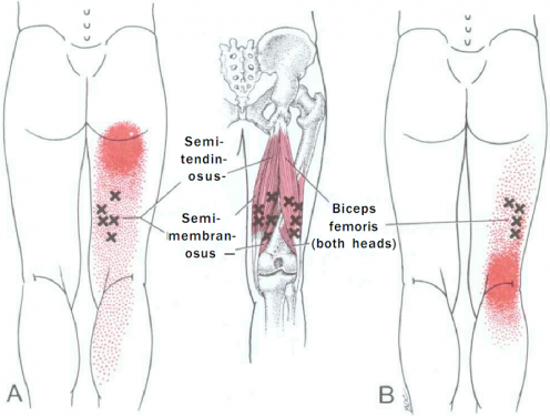
- Pain referred from TrPs in the lower half of the biceps femoris (long or short head) focuses on the back of the knee and may extend up the posterolateral area of the thigh as far as the crease of the buttock.[8]
Treatment[edit | edit source]
Stretching[edit | edit source]
- Position of patient is side lying and support is provided to lower leg with hip and knee flexed[9]
- Hold the upper leg and bring into abduction[9]
- During abduction, try to support the leg against your body, so that the patient can extend the knee whilst pressing the hip into flexion[9]
- With one hand, hold the upper leg and with thenar of your other hand try to provide stretch to the muscle away from the site of insertion[9]
References[edit | edit source]
- ↑ 1.0 1.1 1.2 1.3 http://www.anatomy.tv/interactivehip
- ↑ http://www.primalonlinelearning.com/cedaandp/muscular_system/muscles_of_the_lower_limb.aspx#bicepsfemoris
- ↑ 3.0 3.1 3.2 3.3 3.4 3.5 3.6 3.7 Netter FH (2014). Atlas of Human Anatomy (6th ed). Philadelphia, PA: Saunders-Elsevier.
- ↑ Available from: Kenhub-Learn Human Anatomyhttps://www.youtube.com/watch?v=3qmWlrdNUGA Functions of the biceps femoris muscle-Human 3D anatomy|Kenhub
- ↑ 5.0 5.1 5.2 http://www.wheelessonline.com/ortho/biceps_femoris
- ↑ 6.0 6.1 6.2 6.3 Reichert B, Stelzenmueller W. Palpation Techniques: Surface Anatomy for Physical Therapists. Thieme; New York. 2011.
- ↑ 7.0 7.1 7.2 7.3 Sanzo P, MacHutchon M (2015). Length Tension Testing Book 1, Lower Quadrant: A Workbook of Manual Therapy Techniques (2nd ed). Canada: Brush Education.
- ↑ Travell JG, Simons DG, Simons LS (1998). Travell and Simons' Myofascial Pain and Dysfunction: The Trigger Point Manual, Volume 1: The Lower Extremities (2nd ed). Baltimore, MD: Williams & Wilkins.
- ↑ 9.0 9.1 9.2 9.3 Jari Ylinen, Leon Chaitow (2008). Stretching therapy for Sports and Manual therapies (1st ed) Medirehabook Inc


