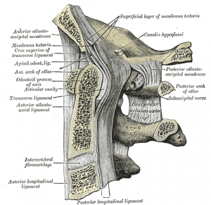
Apical ligament
Original Editor - Rachael Lowe
Top Contributors - Rachael Lowe, Kim Jackson, Evan Thomas, George Prudden and WikiSysop
Description[edit | edit source]
Spans between the second cervical vertebra in the neck and the skull. It lies as a fibrous cord in the triangular interval between the alar ligaments.
Attachments[edit | edit source]
Arises from the apex of the odontoid process on the Axis to the anterior margin of the foramen magnum. It blendes with the deep portion of the anterior atlanto-occipital membrane and superior crus of the Transverse ligament of the atlas.
Function[edit | edit source]
Assists in stabilising the craniocervical junction (head on the vertebral column)[1].
Pathology[edit | edit source]
Examination[edit | edit source]
Recent Related Research (from Pubmed)[edit | edit source]
Failed to load RSS feed from http://www.ncbi.nlm.nih.gov/entrez/eutils/erss.cgi?rss_guid=1P9hIDIGI6NMG3UBOnfBdomWYCgvlgaTyYX0WHFrem_nJ0lYOb|charset=UTF-8|short|max=10: Error parsing XML for RSS
References[edit | edit source]
- ↑ Tubbs RS, Grabb P, Spooner A, Wilson W, Oakes WJ. The apical ligament: anatomy and functional significanceJ Neurosurg. 2000 Apr;92(2 Suppl):197-200.


