Anterior Cruciate Ligament (ACL): Difference between revisions
Rujuta Naik (talk | contribs) mNo edit summary |
Rujuta Naik (talk | contribs) mNo edit summary |
||
| Line 99: | Line 99: | ||
|} | |} | ||
</div> | </div> | ||
Revision as of 14:09, 20 July 2023
Original Editor - Rachael Lowe
Top Contributors - Laura Ritchie, Admin, Kim Jackson, Rujuta Naik, Evan Thomas, Bruno Serra, Joao Costa, 127.0.0.1, Scott Buxton, George Prudden, WikiSysop, Vidya Acharya, Rucha Gadgil and Ewa Jaraczewska Tarang Jai
Introduction[edit | edit source]
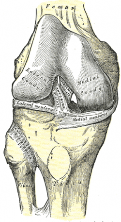
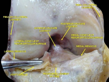
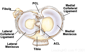
The anterior cruciate ligament (ACL) is a band of dense connective tissue which courses from the femur to the tibia. The ACL is a key structure in the knee joint, as it resists anterior tibial translation and rotational loads.
|
|
Attachments[edit | edit source]
Origin[edit | edit source]
Arises from the posteromedial corner of medial aspect of lateral femoral condyle in the intercondylar notch[1]. This femoral attachment of ACL is on posterior part of medial surface of lateral condyle well posterior to longitudinal axis of the femoral shaft. The attachment is actually an interdigitation of collagen fibers & rigid bone thru transitional zone of
fibrocartilage and mineralized fibrocartilage[2].
Orientation[edit | edit source]
It runs inferiorly, medially and anteriorly.
Insertion[edit | edit source]
It inserts anterior to the inter-condyloid eminence of the tibia, blending with the anterior horn of the medial meniscus. The tibial attachment is in a fossa in front of and lateral to the anterior spine, a rather wide area from 11 mm in width to 17 mm in AP direction[1].
For more detail on the anatomy of the ACL, please see this page: Anterior Cruciate Ligament (ACL) - Structure and Biomechanical Properties
Nerve Supply[edit | edit source]
The ACL receives nerve fibres from the posterior articular branches of the tibial nerve. [3] These fibres penetrate the posterior joint capsule and run along with the synovial and periligamentous vessels surrounding the ligament to reach as far anterior to the infrapatellar fat pad.[3] Most of the fibres are associated with the endoligamentous vasculature and have a vasomotor function. The receptors of the nerve fibres mentioned are as follows:
- Ruffini receptors are sensitive to stretching and are located at the surface of the ligament, predominantly on the femoral portion where the deformations are the greatest. [4]
- Vater–Pacini receptors are sensitive to rapid movements and are located at the femoral and tibial ends of the ACL. [4]
- Golgi-like tension receptors are located near the attachments of the ACL as well as at its surface, beneath the synovial membrane. [3]
- Free-nerve endings function as nociceptors, but they may also serve as local effectors by releasing neuropeptides with vasoactive function. Thus, they may have a modulatory effect in normal tissue homeostasis or in the late remodelling of grafts. [4][5]
The mechanoreceptors cited above (Ruffini, Pacini, and Golgi-like receptors) have a proprioceptive function and provide the afferent arc for signalling knee postural changes. Deformations within the ligament influence the output of muscle spindles through the fusimotor system. [5] Hence, activation of afferent nerve fibres in the proximal part of the ACL influences motor activity in the muscles around the knee; a phenomenon called the ‘‘ACL reflex.’’ These muscular responses are elicited by stimulation of group II or III fibres (i.e. mechanoreceptors). The ACL reflex is an essential part of normal knee function and is involved in the updating of muscle programs.[6] This becomes even more obvious in patients with a ruptured ACL, where the loss of feedback from mechanoreceptors in the ACL leads to quadriceps femoris weakness.[6] Indeed, this afferent feedback from the ACL has a major influence on the maximal voluntary contraction exertion of the quadriceps femoris. [7]
Vascular Supply[edit | edit source]
| [8] |
The major blood supply of the cruciate ligaments arises from the middle geniculate artery.[9] [10] The distal part of both cruciate ligaments is vascularized by branches of the lateral and medial inferior geniculate artery.[10] The ligament is surrounded by a synovial fold where the terminal branches of the middle and inferior arteries form a periligamentous network. From the synovial sheath blood vessels penetrate the ligament in a horizontal direction and anastomose with a longitudinally orientated intraligamentous vascular network.[11] The density of blood vessels within the ligaments is not homogeneous.[12] In the ACL, an avascular zone is located within the fibrocartilage of the anterior part where the ligament faces the anterior rim of the intercondylar fossa.[12] The coincidence of poor vascularity and the presence of fibrocartilage is also seen in gliding tendons in areas that are subjected to compressive loads, and the coincidence of these two factors undoubtedly plays a role in the poor healing potential of the ACL. [13]
Composition[edit | edit source]
The ACL has a microstructure of collagen bundles of multiple types (mostly type I) and a matrix made of a network of proteins, glycoproteins, elastic systems, and glycosaminoglycans with multiple functional interactions[14].
Bundles[edit | edit source]
| [15] |
There are three components of the ACL, the smaller anteromedial bundle (AMB) , the intermediate bundle and the larger posterolateral bundle (PLB), named according to where the bundles insert into the tibial plateau.[16]
The anteromedial bundle is tight in flexion whereas the posterolateral bundle is stretched in extension. In extension both bundles are parallel. In flexion, the femoral insertion site of the posterolateral bundle moves anteriorly, both bundles are crossed, the anteromedial bundle tightens and the posterolateral bundle loosens.
With the knee extended, resistance to anterior translation of the tibia, Lachmans Test, is by the bulky posterolateral bundle. With the knee flexed, resistance to anterior translation of the tibia, the Anterior Drawer Test, is by the anterior medial bundle.
Rupture of the posterolateral bundle causes an increase in hyperextension, anterior translation (extended knee), increase in external and internal rotation (knee extended), and increases in external rotation with the knee in mid-flexion; Rupture of the anteromedial bundle causes anterolateral instability with an increase in anterior translation in flexion, minimal increase in hyperextension, and minimal rotational instability.
For more detail on the ACL bundles, please see this page: Anterior Cruciate Ligament (ACL) - Structure and Biomechanical Properties
Function[edit | edit source]
The ACL provides approximately 85% of the total restraining force of anterior translation. It also prevents excessive tibial medial and lateral rotation, as well as varus and valgus stresses. To a lesser degree, the ACL checks extension and hyperextension. Together with the posterior cruciate ligament (PCL), the ACL guides the instantaneous center of rotation of the knee, therefore controlling joint kinematics. While the anteromedial bundle is the primary restraint against anterior tibial translation, the posterolateral bundle tends to stabilize the knee near full extension, particularly against rotatory loads[17].
Video[edit | edit source]
Presentations[edit | edit source]
 |
Anterior Cruciate Ligament Injury
This presentation, created by Terdsak Rojsurakitti, Doctor at Managed Care, discusses anatomy, mechanism of injury, surgical options and rehabilitation of ACL tears. Anterior Cruciate Ligament Injury/ View the presentation |
References[edit | edit source]
- ↑ 1.0 1.1 Mark L. Purnell, Andrew I. Larson, and William Clancy. Anterior Cruciate Ligament Insertions on the Tibia and Femur and Their Relationships to Critical Bony Landmarks Using High-Resolution Volume-Rendering Computed Tomography. Am J Sports Med November 2008 vol. 36 no. 11 2083-2090
- ↑ Wheeless, C,R. Wheeless' Textbook of Orthopaedics. http://www.wheelessonline.com/ortho/anatomy_of_acl Accessed 8/1/12.
- ↑ 3.0 3.1 3.2 Kennedy JC, Alexander IJ, Hayes KC. Nerve supply of the human knee and its functional importance. Am J Sports Med. 1982 Nov-Dec;10(6):329-35.
- ↑ 4.0 4.1 4.2 Haus J, Halata Z. Innervation of the anterior cruciate ligament. Int Orthop. 1990;14(3):293-6.
- ↑ 5.0 5.1 Hogervorst T, Brand RA. Mechanoreceptors in joint function. J Bone Joint Surg Am. 1998 Sep;80(9):1365-78.
- ↑ 6.0 6.1 Konishi Y, Fukubayashi T, Takeshita D. Possible mechanism of quadriceps femoris weakness in patients with ruptured anterior cruciate ligament. Med Sci Sports Exerc. 2002 Sep;34(9):1414-8.
- ↑ Konishi Y, Suzuki Y, Hirose N, Fukubayashi T. Effects of lidocaine into knee on QF strength and EMG in patients with ACL lesion. Med Sci Sports Exerc. 2003 Nov;35(11):1805-8.
- ↑ Dr. Bertram Zarins, MD. Knee Ligament Anatomy Animation. Available from: http://www.youtube.com/watch?v=RTV5Yo3E7VQ[last accessed 04/10/14]
- ↑ Arnoczky SP. Anatomy of the anterior cruciate ligament. Clin Orthop Relat Res. 1983 Jan-Feb;(172):19-25.
- ↑ 10.0 10.1 Scapinelli R. Vascular anatomy of the human cruciate ligaments and surrounding structures. Clin Anat. 1997;10(3):151-62.
- ↑ Arnoczky SP, Rubin RM, Marshall JL. Microvasculature of the cruciate ligaments and its response to injury. An experimental study in dogs. J Bone Joint Surg Am. 1979 Dec;61(8):1221-9.
- ↑ 12.0 12.1 Petersen W, Tillmann B. Structure and vascularization of the cruciate ligaments of the human knee joint. Anat Embryol (Berl). 1999 Sep;200(3):325-34.
- ↑ Giori NJ, Beaupré GS, Carter DR. Cellular shape and pressure may mediate mechanical control of tissue composition in tendons. J Orthop Res. 1993 Jul;11(4):581-91.
- ↑ Duthon VB, Barea C, Abrassart S, Fasel JH, Fritschy D, Ménétrey J. Anatomy of the anterior cruciate ligament. Knee Surg Sports Traumatol Arthrosc. 2006 Mar;14(3):204-13. Epub 2005 Oct 19.
- ↑ Non-Contact ACL Injury, Treatment and Rehabilitation Animation. The Normal Function of the Anterior Cruciate Ligament. Available from: http://www.youtube.com/watch?v=RwwxtD-xT4Y[last accessed 04/10/14]
- ↑ Amis AA, Dawkins GP. Functional anatomy of the anterior cruciate ligament. Fibre bundle actions related to ligament replacements and injuries. The Journal of Bone & Joint Surgery British Volume. 1991 Mar 1;73(2):260-7.
- ↑ Petersen W, Zantop T. Anatomy of the anterior cruciate ligament with regard to its two bundles. Clin Orthop Relat Res. 2007 Jan;454:35-47


