Anatomy of the Human Heart: Difference between revisions
No edit summary |
No edit summary |
||
| Line 65: | Line 65: | ||
== Conducting System of Heart == | == Conducting System of Heart == | ||
[[File:Heart conduction system .jpeg|right|frameless|708x708px]] | |||
An electrical conduction system regulates the pumping of the heart and timing of contraction of various chambers. Heart muscle contracts in response to the electrical stimulus received system generates electrical impulses and conducts them throughout the muscle of the heart, stimulating the heart to contract and pump blood. Among the major elements in the cardiac conduction system are the sinus node, atrioventricular node, and the autonomic nervous system. <ref name=":0" /> | An electrical conduction system regulates the pumping of the heart and timing of contraction of various chambers. Heart muscle contracts in response to the electrical stimulus received system generates electrical impulses and conducts them throughout the muscle of the heart, stimulating the heart to contract and pump blood. Among the major elements in the cardiac conduction system are the sinus node, atrioventricular node, and the autonomic nervous system. <ref name=":0" /> | ||
| Line 95: | Line 96: | ||
== Nerve Supply == | == Nerve Supply == | ||
The heart | The main control of the heart resides with the medulla oblongata. There is an area called the cardioacceleratory centre, or pressor centre, in the upper part of the medulla oblongata, and an area called the cardioinhibitory centre, or depressor centre, in the lower part. Together they are called the cardioregulatory centre, since they interact to control heart rate, etc. | ||
The | The nervous supply to the heart is autonomic, consisting of both sympathetic and parasympathetic parts. The sympathetic fibres arise from the pressor centre, while the parasympathetic fibres arise in the depressor centre. | ||
The | The sympathetic nervous system acts on the sinoatrial node, speeding up the depolarisation rate, and therefore increasing the heart rate. The parasympathetic system works in reverse in order to slow the heart rate down. The heart itself has a natural pacemaker, the sinoatrial node, which does not need a nervous supply to function. If you sever all the nerves to the heart, then it will continue to beat. In fact, it will beat faster than normal, since there is normally a parasympathetic supply slowing the heart down.<ref>Basic heart Anatomy Nervous Supply Heart Available: https://www.liverpool.ac.uk/~trh/local_html/heartdisease/nerve_supply_to_the_heart.htm (accessed18.7.2021)</ref> | ||
== Relevance to Physiotherapy == | == Relevance to Physiotherapy == | ||
Revision as of 01:03, 18 July 2021
Original Editor - Uchechukwu Chukwuemeka
Top Contributors - Uchechukwu Chukwuemeka, Lucinda hampton, Kim Jackson, George Prudden, Joao Costa, Sai Kripa, Admin and Adam Vallely Farrell
Introduction[edit | edit source]

The heart is a muscular organ that serves to collect deoxygenated blood from all parts of the body, carries it to the lungs to be oxygenated and release carbon dioxide. Then, it transports the oxygenated blood from the lungs and distributes it to all the body parts[1]
- The heart pumps around 7,200 litres of blood in a day throughout the body[2].
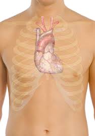
- The heart is situated at the centre of the chest and points slightly towards the left.
- On average, the heart beats about 100,000 times a day, i.e., around 3 billion beats in a lifetime.
- An adult heart beats about 60 to 80 times per minute, and newborn babies heart beats faster than an adult which is about 70 to 190 beats per minute.

Anatomy[edit | edit source]

The heart has a somewhat conical form and is enclosed by the pericardium. It is positioned posteriorly to the body of the sternum with one-third situated on the right and two-thirds on the left of the midline. The heart measures 12 x 8.5 x 6 cm and weighs ~310 g (males) and ~255 g (females)
Relations
- Anteriorly: the body of the sternum, and adjoining costal cartilages; left lung, and pleura (apex)
- Posteriorly: oesophagus, descending thoracic aorta, azygos, hemiazygos veins, and thoracic duct
- Superficially : bifurcation of the main pulmonary trunk
- Inferiorly: diaphragm
- Laterally: lungs, pleura[3]
Layers of the Heart Walls[edit | edit source]

The heart wall consists of three layers enclosed in the pericardium[1][4][5]:
- Epicardium - the outer layer of the wall of the heart and is formed by the visceral layer of the serous pericardium.
- Myocardium - the muscular middle layer of the wall of the heart and has excitable tissue and the conducting system.
- Endocardium .[3]
- A middle concentric layer
- A subendocardial layer.
The rest of the heart is composed mainly of the subepicardial and subendocardial layers.
Structure and Function[edit | edit source]

The heart is subdivided by septa into right and left halves, and a constriction subdivides each half of the organ into two cavities, the upper cavity being called the atrium, the lower the ventricle. The heart, therefore, consists of four chambers:
- right atrium
- left atrium
- right ventricle
- left ventricle[3].
It is best to remember the four chambers and four valves in order of the series that blood travels through the heart:
- Venous blood returning from the body drains into the right atrium via the SVC, IVC and coronary sinus
- The right atrium pumps blood through the tricuspid valve into the right ventricle
- The right ventricle pumps blood through the pulmonary semilunar valve into the pulmonary trunk to be oxygenated in the lungs
- Blood returning from the lungs drains into the left atrium via the four pulmonary veins
- The left atrium pumps blood through the bicuspid (mitral) valve into the left ventricle
- The left ventricle pumps blood through the aortic semilunar valve into the ascending aorta to supply the body.[3]
Heart Valves[edit | edit source]

The heart has the Four valves. All four valves of the heart have a singular purpose: allowing forward flow of blood but preventing backward flow.[6]
- Pulmonary Valve - The opening into the pulmonary trunk is closed by the pulmonary valve, which consists of three semilunar cusps with free edges projecting upward into the lumen of the pulmonary trunk thus prevents the backflow of blood as it is pumped from the right ventricle to the pulmonary artery. The cusps are named the left, right and anterior semilunar cusps, relative to their fetal position. Each cusp forms a pocket-like sinus; a dilation in the wall of the initial portion of the pulmonary trunk.[5]
- Tricuspid Valve - The right atrioventricular opening is closed during ventricular contraction by the tricuspid valve (right atrioventricular valve) thus prevents the backflow of blood as it is pumped from the right atrium to the right ventricle. It’s named tricuspid valve because it made up of three cusps or leaflets. The naming of the three cusps, the anterior, septal, and posterior cusps, is based on their position in relation to the right ventricle.
- Aortic Valve - The opening from the left ventricle into the aorta is closed by the aortic valve thus prevents the backflow of blood as it is pumped from the left ventricle to the aorta. It consists of three semilunar cusps with the free edge of each projecting upward into the lumen of the ascending aorta. Between the semilunar cusps and the wall of the ascending aorta are pocket-like sinuses-the right, left, and posterior aortic sinuses. The right and left coronary arteries emanates from the right and left aortic sinuses; Thus, the posterior aortic sinus and cusp are sometimes called noncoronary sinus and cusp.
- Mitral Valve - The left atrioventricular orifice opens into the posterior right side of the superior part of the left ventricle. It is closed during ventricular contraction by the mitral valve (left atrioventricular valve), which is also referred to as the bicuspid valve as it has two cusps; the anterior and posterior cusps. It prevents the backflow of blood as it is pumped from the left atrium to the left ventricle and are continuous with each other at the commissures.[5][7]
Conducting System of Heart[edit | edit source]

An electrical conduction system regulates the pumping of the heart and timing of contraction of various chambers. Heart muscle contracts in response to the electrical stimulus received system generates electrical impulses and conducts them throughout the muscle of the heart, stimulating the heart to contract and pump blood. Among the major elements in the cardiac conduction system are the sinus node, atrioventricular node, and the autonomic nervous system. [6]
- The sinus node is the heart's natural pacemaker. The sinus node is a cluster of cells situated in the upper part of the wall of the right atrium. The electrical impulses are generated there. (The sinus node is also called the sinoatrial node.)
- The electrical signal generated by the sinus node moves from cell to cell down through the heart until it reaches the atrioventricular node (the AV node), a cluster of cells situated in the center of the heart between the atria and ventricles.
- The AV node serves as a gate that slows the electrical current before the signal is permitted to pass down through to the ventricles. This delay ensures that the atria have a chance to fully contract before the ventricles are stimulated. After passing the AV node, the electrical current travels to the ventricles along special fibers embedded in the walls of the lower part of the heart.
- The autonomic nervous system (the same part of the nervous system as controls the blood pressure) controls the firing of the sinus node to trigger the start of the cardiac cycle. The autonomic nervous system can transmit a message quickly to the sinus node so it in turn can increase the heart rate to twice normal within only 3 to 5 seconds. This quick response is important during exercise when the heart has to increase its beating speed to keep up with the body's increased demand for oxygen.[8]
Blood Supply[edit | edit source]
Image: Coronary Artery Territories

The heart is supplied by two coronary arteries:
- Left main coronary artery carries 80% of the flow to the heart muscle. It is a short artery that divides into two branches
- Left anterior descending artery that supplies anterior two-thirds of the inter-ventricular septum and adjoining part of the left ventricular anterior wall
- Circumflex coronary artery that supplies blood to the lateral and posterior portions of the left ventricle.
2. Right coronary artery: branches supply the right ventricle, right atrium, and left ventricle's inferior wall.
Coronary arteries and veins course over the surface of the heart. Most coronary veins coalesce into the coronary sinus that runs in the left posterior atrioventricular groove and opens into the right atrium. Other small veins, called thebesian veins, open directly into all four chambers of the heart.[6]
Venous drainage[edit | edit source]
Venous drainage is via the variable coronary veins and the coronary sinus[3].
Lymphatic Drainage of the Heart[edit | edit source]
The lymphatic vessels drain mainly into:
- Brachiocephalic nodes, in front of brachiocephalic veins
- Tracheobronchial nodes, located at the distal end of the trachea[7].[5]
Nerve Supply[edit | edit source]
The main control of the heart resides with the medulla oblongata. There is an area called the cardioacceleratory centre, or pressor centre, in the upper part of the medulla oblongata, and an area called the cardioinhibitory centre, or depressor centre, in the lower part. Together they are called the cardioregulatory centre, since they interact to control heart rate, etc.
The nervous supply to the heart is autonomic, consisting of both sympathetic and parasympathetic parts. The sympathetic fibres arise from the pressor centre, while the parasympathetic fibres arise in the depressor centre.
The sympathetic nervous system acts on the sinoatrial node, speeding up the depolarisation rate, and therefore increasing the heart rate. The parasympathetic system works in reverse in order to slow the heart rate down. The heart itself has a natural pacemaker, the sinoatrial node, which does not need a nervous supply to function. If you sever all the nerves to the heart, then it will continue to beat. In fact, it will beat faster than normal, since there is normally a parasympathetic supply slowing the heart down.[9]
Relevance to Physiotherapy[edit | edit source]
- The surface markings of the heart and the position of the apex beat can show if the heart has shifted its position in relation to the chest wall or if the heart is enlarged by disease. The position of the margins of the heart can be determined by percussion (Snell, 2000).
- Percussion defines the density and size of the heart. It is performed at the 3rd, 4th, and 5th intercostal spaces from the left anterior axillary line to the right anterior axillary line.
- The pericardial sac is influenced by movements of the heart and great vessel, sternum, and diaphragm. The heart moves downwards on deep inspiration since the heart’s position depends on that of the diaphragm.
- The fibrous pericardium protects the heart against sudden overfilling and anchors it within the mediastinum.
- If a coronary artery becomes obstructed, by a blood clot for example, part of the myocardium becomes ischemic, that is, deprived of its blood supply. Prolonged ischemia will create an infarct, an area of necrotic (dead) tissue. This is a myocardial infarction, commonly called a heart attack (myocardial infarction).

Also relevant to physiotherapy: Acute Coronary Syndrome; Physical Activity and Cardiovascular Disease; Cardiac Rehabilitation; Myocardial Infarction; Atrial Fibrillation.
Videos[edit | edit source]
References[edit | edit source]
- ↑ 1.0 1.1 Kenny, WL,Wilmore, JH, Costill, DL. Cardiovascular System and its Control. In Physiology of Sport and Exercise, 5rdedn. Human Kinetics, 2011. 140-150.
- ↑ Inovqheart Heart Available from: https://www.inovaheart.org/upload/docs/Healthcare%20Services/Heart%20and%20Vascular/fast-facts-about-the-heart.pdf
- ↑ 3.0 3.1 3.2 3.3 3.4 Radiopedia Heart Available: https://radiopaedia.org/articles/heart?lang=gb (accessed 17.6.2021)
- ↑ Malouf, JF, Edwards, WD, Tajil, AJ, Seward, JB. Functional anatomy of the heart. In: Fuster, F, Alexander, RW, O’Rourke, RA editors. Hurst’s: The Heart. 10th edn. McGraw-Hill Inc., 2001. p19–62.
- ↑ 5.0 5.1 5.2 5.3 Drake, RL, Vogl, W, Mitchell, AW, Gray, H. Gray's anatomy for Students 2nd ed. Philadelphia : Churchill Livingstone/Elsevier, 2010.
- ↑ 6.0 6.1 6.2 Rehman I, Rehman A. Anatomy, Thorax, Heart. [Updated 2020 Dec 28]. In: StatPearls [Internet]. Treasure Island (FL): StatPearls Publishing; 2021 Jan Available:https://www.ncbi.nlm.nih.gov/books/NBK470256/ (accessed 17.6.2021)
- ↑ 7.0 7.1 Moore, KL, Dalley, AF. Clinically oriented anatomy. 6th ed. Philadelphia: Lippincott Williams & Wilkins. 2009
- ↑ Medicine net Conduction system heart Available:https://www.medicinenet.com/heart_conduction_system/definition.htm (accessed 18.7.2021)
- ↑ Basic heart Anatomy Nervous Supply Heart Available: https://www.liverpool.ac.uk/~trh/local_html/heartdisease/nerve_supply_to_the_heart.htm (accessed18.7.2021)

