Alar ligaments: Difference between revisions
mNo edit summary |
mNo edit summary |
||
| Line 31: | Line 31: | ||
== Recent Related Research (from [http://www.ncbi.nlm.nih.gov/pubmed/ Pubmed]) == | == Recent Related Research (from [http://www.ncbi.nlm.nih.gov/pubmed/ Pubmed]) == | ||
<div class="researchbox"> | <div class="researchbox"> | ||
<rss>http://www.ncbi.nlm.nih.gov/entrez/eutils/erss.cgi?rss_guid=1pcFRJI0caoCrdhC_H3wm_dPxs0fILo8zPCFvz5WO | <rss>http://www.ncbi.nlm.nih.gov/entrez/eutils/erss.cgi?rss_guid=1pcFRJI0caoCrdhC_H3wm_dPxs0fILo8zPCFvz5WO</rss> | ||
</div> | </div> | ||
== References == | == References == | ||
Revision as of 08:25, 7 June 2017
Original Editor - Rachael Lowe
Top Contributors - Rachael Lowe, Kim Jackson, Evan Thomas, WikiSysop, Alistair James, George Prudden and Wendy Snyders
Description[edit | edit source]
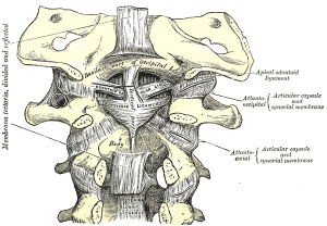
Two strong rounded cords that attach the skull to C2 (Axis).
Attachments[edit | edit source]
Arise from either side of the odontoid process and attach to the medial aspect of the occipital condyles.
Function[edit | edit source]
Taut in flexion, limit rotation and side flexion to the opposite side.
Play a role in stabilizing C1 and C2, especially in rotation[1].
Pathology[edit | edit source]
Injured in rear end motor vehicle collisions when the cervical spine is in extremes of rotation.
Examination
[edit | edit source]
Recent Related Research (from Pubmed)[edit | edit source]
Failed to load RSS feed from http://www.ncbi.nlm.nih.gov/entrez/eutils/erss.cgi?rss_guid=1pcFRJI0caoCrdhC_H3wm_dPxs0fILo8zPCFvz5WO: Error parsing XML for RSS
References[edit | edit source]
- ↑ Magee DJ (2007). Orthopedic Physical Assessment (5th ed). St Louis, MO: Saunders Elsevier.


