Abdominal Muscles: Difference between revisions
No edit summary |
No edit summary |
||
| Line 1: | Line 1: | ||
<div class="noeditbox"> | |||
This article or area is currently under construction and may only be partially complete. It will be changed to describe anterior and posterior abdominal walls. Please come back soon to see the finished work! ({{REVISIONDAY}}/{{REVISIONMONTH}}/{{REVISIONYEAR}}) | |||
</div> | |||
<div class="editorbox"> | <div class="editorbox"> | ||
'''Original Editor ''' - [[User:Anne Millar|Anne Millar]] | '''Original Editor ''' - [[User:Anne Millar|Anne Millar]] | ||
Revision as of 01:58, 14 May 2020
This article or area is currently under construction and may only be partially complete. It will be changed to describe anterior and posterior abdominal walls. Please come back soon to see the finished work! (14/05/2020)
Original Editor - Anne Millar
Top Contributors - Anne Millar, Khloud Shreif, Lucinda hampton, Laura Ritchie, Kim Jackson, Admin, Joao Costa, Scott Buxton, Vidhu Sindwani and Evan Thomas
Introduction[edit | edit source]
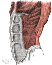
The abdominal muscles form of the anterolateral and anterior abdominal wall. The anterolateral abdominal wall consists of three flat muscles their fibers run in different directions overlap each other. consists of external abdominal oblique the most superficial one directed inferiorly medially, internal abdominal oblique intermediate muscle directed superior medially perpendicular to external oblique. And transversus abdominis its fibers run transversely like the front of a belt on pants. This overlap supports the abdominal wall and decreases the risk of abdominal herniation. The anterior wall consists of two vertical muscles located on the midline and bisected by linea alba; Rectus abdominis and pyramidalis. Acting together these muscles form a firm wall that supports the muscles of the spine and help to maintain an erect posture, Support internal visceral organs where there is no bone. In addition the contraction of these muscles helps in forceful expiration and to increases the intra-abdominal pressure such as in sneezing, coughing, micturating, defecating, lifting, and childbirth.[1]
External Abdominal Oblique[edit | edit source]
Structure[edit | edit source]
The external abdominal oblique muscle is the largest and most superficial of the four muscles and lies on the sides and front of the abdomen. It is broad and thin with it's muscular portion occupying the side and it's aponeurosis the anterior wall of the abdomen.
Origin: it arises from the external surface and inferior borders of the lower eight ribs. The fibres from the lowest ribs pass nearly vertically downwards.
Insertion: the lower fibres inserted into the anterior half of outer lip the iliac crest, the middle and upper fibres, directed inferiorly and anteriorly, end in an aponeurosis at approximately the mid-clavicular line (that aponeurosis form inguinal ligament) and insert into the linea alba, the pubic crest and the pubic tubercle [2].
Innervation[edit | edit source]
Nerve supply: the upper two-third of external abdominal oblique supplied by the lower six thoracic nervesT7-T11, the lower third supplied by the iliohypogastricL1 and ilioinguinal nerves[3].
Blood supply: the upper 2/3 receive blood supply from branches of lower posterior intercostal and subcostal arteries, lower 1/3 from deep circumflex iliac artery.[4]
Function [edit | edit source]
Both sides acting together, the external abdominal obliques flex the vertebral column by drawing the pubis towards the xiphoid process. Acting unilaterally it results in ipsilateral side flexion and contralateral rotation of the trunk.[5]
Internal Abdominal Oblique[edit | edit source]
Structure[edit | edit source]
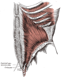
The internal abdominal oblique muscle is also a broad thin muscular sheet that lies deep to the external oblique muscle.
Origin: it arises from the thoraco-lumbar fascia, the anterior two-thirds of the iliac crest and the lateral two-thirds of the inguinal ligament.
Insertion: the muscle fibres radiate superomedially and insert into the inferior borders of the lower three ribs and their costal cartilages, the xiphoid process, the linea alba and the symphysis pubis.
Near their insertion the lowest tendinous fibres are joined with similar fibres from the transversus abdominis to form the conjoint tendon[2].[6] [7]
Innervation[edit | edit source]
Nerve supply: the internal abdominal obliques are innervated by the lower six thoracic nerves and the iliohypogastric and ilioinguinal nerves[3].
Blood supply: lower posterior intercostal and subcostal arteries, superior and inferior epigastric arteries, superficial and deep circumflex iliac arteries, posterior lumbar arteries.
Function[edit | edit source]
Acting unilaterally, contraction of the internal oblique results in ipsilateral side flexion and rotation of the trunk.[5] It acts with the external oblique muscle of the opposite side to achieve this torsional movement of the trunk. It also acts to compress the abdominal viscera, pushing them up into the diaphragm, resulting in a forced expiration.
Transversus Abdominis[edit | edit source]
Structure[edit | edit source]
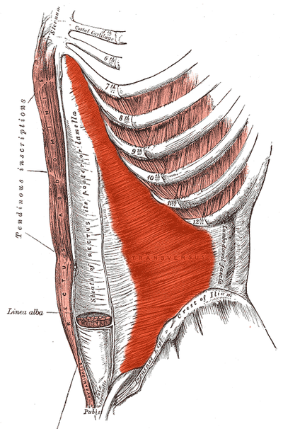
The transversus abdominis muscle is the deepest of the abdominal muscles, lying internally to the internal abdominal obliques. It is a thin sheet of muscle whose fibres run transversely anteriorly perpendicular to linea alba. it frames the lateral abdominal muscle
Origin: it arises as fleshy fibres from the deep surface of the lower six costal cartilages, the lumbar fascia, the anteror two-thirds of the iliac crest and the lateral third of the inguinal ligament.
Insertion: it inserts into the xiphoid process, the linea alba and the symphsis pubis. The lowest tendinous fibres join similar fibres from the interior obliques to form the conjoint tendon which is fixed to the pubic crest and the pectineal line[2].[8]
Innervation[edit | edit source]
Nerve supply: the transversus abdominus muscle is innervated by the lower six thoracic nerves and the iliohypogastric L1 and ilioinguinal nerves L1[3].
Blood supply: same as internal abdominal oblique; lower posterior intercostal and subcostal arteries, superior and inferior epigastric arteries, superficial and deep circumflex iliac arteries, posterior lumbar arteries.
Function[edit | edit source]
Contraction of the transversus abdominis has a corset like effect, narrowing and flattening the abdomen. Its primary function is to stabilise the lumbar spine and pelvis before movement of the lower and /or upper limbs occur[9].
Rectus Abdominis[edit | edit source]
Structure[edit | edit source]
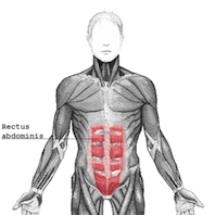
The rectus abdominis is a long strap muscle that extends the entire length of the anterior abdominal wall. It is broader above and lies close to the midline, being separated from it's fellow by the linea alba.
Origin: it arises by two heads from the anterior aspect of the symphysis pubis and the pubic crest.
Insertion: it inserts into the 5th , 6th and 7th costal cartilages and the xiphoid process.
Each rectus abdominis is divided into three distinct segments by three transverse fibrous intersections, one at the level of the xiphoid process, one at the umbilicus and one halfway between the two. The rectus abdominis is enclosed between the aponeuroses of the external and internal obliques and transversus abdominis which form the rectus sheath[2].[10]
Innervation[edit | edit source]
Nerve supply: the rectus abdominis muscle is innervated by the lower six thoracic nerves[3].
Blood supply: superior and inferior epigastric arteries; inferior is the main supply of rectus abdominis[11]
Function[edit | edit source]
The rectus abdominis is an important postural and core muscle . With a fixed pelvis, contraction results in flexion of the lumbar spine. When the ribcage is fixed contraction results in a posterior pelvic tilt. It also plays an important role in forced expiration and in increasing intra-abdominal pressure.[5]
| [12] |
Pyramidalis[edit | edit source]
Structure[13][edit | edit source]
Pyramidalis with rectus abdominis contribute to form the anterior abdominal wall. It's absent in 20% of the population and has less significant role, triangular muscle.
Origin: it arises from symphysis pubic and pubic crest
Insertion: decrease in size as it ascends and inserts medially to linea alba as apointed apex.
Innervation[edit | edit source]
Nerve supply: It is innervated by subcostal nerve T12
Blood supply: the main arterial supply from the inferior epigastric supply and the deep circumflex iliac artery to a lesser extent.
Function[edit | edit source]
When they contract tense the linea alba
Clinical relevance[edit | edit source]
Transverse abdomimis as a deep abdominal muscle and one of main important core muscle that contributes to supporting lumbopelvic stability and deficit in its function affect our back causing low back pain (LBP). We need to include it in our rehabilitation program[14]
Deficit in the abdominal wall muscles congenital from birth or acquired postoperatively as a result of a poor wound healing, wound infection, or acquired weakness after pregnancy and labor for example. can manifest in the form of hernia congenital or acquired.
Congenital hernia happens during infant development as a result of embryological malformations or weakness in neonatal abdominal wall, it may be fatal in some cases and need urgent and surgical intervention as; gastroschisis, or resolve without need to surgical intervention as in umbilical hernia.
Acquired hernia happens in the area of weakness and varies in its severity[15]:
Umbilical hernia that is more serious and has a higher rate of morbidity in adult more than infants and may need surgical intervention. Inguinal hernia protrudes at the inferior border of anterolateral muscles. Epigastric hernia, above the umbilicus through the midline of linea alba. Spigelian hernias, and incisional hernia as a result of postoperative incision.
Rectus diastasis happens due to prolong transverse stress on linea alba during pregnancy, or post-menopausal women.
physical therapy intervention[edit | edit source]
Abdominal exercises need to be gradually progressed from how to activate muscles and maintain contraction to integrate them with functional movement, but there's special considerations, precaution, and exercise modification that we will take these exercises for a patient with a hernia.
- Abdominal draw in exercise, easy to apply, target mainly transversus abdominis as well as the diaphragm it's an important respiratory exercise[16]. Exercise can be progressed by adding external resistance, upper limb or lower limb movement while holding abdomen drawing in
- Curl up exercise, target rectus abdominis, transverse abdominis, and obliques in addition to hip flexors, chest, and neck, start exercise with slow movement, few repetitions and make sure the back is in contact with the floor.
- Bridging, modified bridging with hip abduction or unstable surface show to increase core stability, trunk control. The activation of internal abdominis, rectus abdominis along with erector spine is greater in modified bridging when compared to standard bridging[18].
- Blank and pilates exercises activate and strengthen core muscles and abdominal muscles
Resources[edit | edit source]
Axial muscles of the abdominal wall and, thorax
TeachMeAnatomy: the anterolateral abdominal wall
Curl up exercise prescription Healthline
For more exercises description
Core stability- physiopedia page
Lumbar motor control training- physiopedia page
References[edit | edit source]
- ↑ Drake RL, Vogyl AW, Mitchell AW. Gray's anatomy for students. 3rd edition. Philadelphia: Churchill Livingstion Elsevier; 2015. 282p.
- ↑ 2.0 2.1 2.2 2.3 Gray H. Grays Anatomy. 37th ed. Edinburgh: Churchill Livingstone, 1989.
- ↑ 3.0 3.1 3.2 3.3 Mantle J, Haslam J, Barton S. Physiotherapy in Obstetrics and Gynaecology. 2nd Ed. Edinburgh: Butterworth Heinemann, 2004
- ↑ https://www.kenhub.com/en/library/anatomy/external-abdominal-oblique-muscle
- ↑ 5.0 5.1 5.2 Drake RL, Vogyl AW, Mitchell AW. Gray's anatomy for students. 3rd edition. Philadelphia: Churchill Livingstion Elsevier; 2015. 286p
- ↑ Wikipedia. Internal Abdominal Obliques. http://en.wikipedia.org/wiki/Abdominal_internal_muscle
- ↑ KENHUBhttps://www.kenhub.com/en/library/anatomy/internal-abdominal-oblique-muscle
- ↑ Wikipedia. Transversus Abdominis. http://en.wikipedia.org/wiki/Transversis_abdominis_muscle
- ↑ Lee D. The Pelvic Girdle. 2nd Ed. Edinburgh: Churchill Livingstone, 1999.
- ↑ Wikipedia. Rectus Abdominis. http://en.wikipedia/org.wiki/Rectus_abdominis_muscle
- ↑ https://www.kenhub.com/en/library/anatomy/anterior-abdominal-muscles
- ↑ Anatomy Zone. Muscles of the Anterior Abdominal Wall-3D Anatomy Tutorial. Available from: http://www.youtube.com/watch?v=mvOajxO8mXO [last accessed 11/07/15]
- ↑ https://www.kenhub.com/en/library/anatomy/pyramidalis-muscle
- ↑ Selkow NM, Eck MR, Rivas S. Transversus abdominis activation and timing improves following core stability training: a randomized trial. International journal of sports physical therapy. 2017 Dec;12(7):1048.
- ↑ Flynn W, Vickerton P. Anatomy, Abdomen and Pelvis, Abdominal Wall. InStatPearls [Internet] 2019 Dec 9. StatPearls Publishing.
- ↑ Oh YJ, Park SH, Lee MM. Comparison of Effects of Abdominal Draw-In Lumbar Stabilization Exercises with and without Respiratory Resistance on Women with Low Back Pain: A Randomized Controlled Trial. Medical Science Monitor: International Medical Journal of Experimental and Clinical Research. 2020;26:e921295-1.
- ↑ Health e-University. How to do a Curl Up: Health e-University. Available from: http://www.youtube.com/watch?v=lsWQ0XpiNkE[last accessed 25/4/2020
- ↑ Yoon JO, Kang MH, Kim JS, Oh JS. Effect of modified bridge exercise on trunk muscle activity in healthy adults: a cross sectional study. Brazilian journal of physical therapy. 2018 Mar 1;22(2):161-7.
- ↑ Physio Fitness | Physio REHAB | Tim Keeley. Glute Bridges and back pain - Don't flex the spine! | Feat. Tim Keeley | No.70 Physio REHAB. Available from: http://www.youtube.com/watch?v=SwyDMwpcW38[last accessed 25/4/2020


