Neuromyelitis Optica: Difference between revisions
Reem Ramadan (talk | contribs) No edit summary |
Reem Ramadan (talk | contribs) No edit summary |
||
| Line 18: | Line 18: | ||
== Clinical Presentation == | == Clinical Presentation == | ||
Patients with NMO experience attacks that come and go. These attacks vary from mild to severe and can last from days to months ad in some cases these attacks may last and become permanent. The symptoms are broken down into three categories: | Patients with NMO experience attacks that come and go. These attacks vary from mild to severe and can last from days to months ad in some cases these attacks may last and become permanent. The symptoms are broken down into three categories<ref>Jarius S, Wildemann B, Paul F. Neuromyelitis optica: clinical features, immunopathogenesis and treatment. Clinical & Experimental Immunology. 2014 May;176(2):149-64.</ref>: | ||
Optic neuritis occurs due to the inflammation of one or both of the optic nerves. Some of the optic neuritis symptoms include: | Optic neuritis (ON) occurs due to the inflammation of one or both of the optic nerves. Some of the optic neuritis symptoms include<ref>Toosy AT, Mason DF, Miller DH. Optic neuritis. The Lancet Neurology. 2014 Jan 1;13(1):83-99.</ref>: | ||
* Decreased visual acuity | * Decreased visual acuity | ||
* Visual field defects | * Visual field defects | ||
* Color blindness | * Color blindness | ||
* Eye pain | * Eye pain | ||
Myelitis | Transverse Myelitis (TM) occur due to the inflammation of the spinal cord and some of it's symptoms include<ref>Frohman EM, Wingerchuk DM. Transverse myelitis. New England Journal of Medicine. 2010 Aug 5;363(6):564-72.</ref>: | ||
* Muscle weakness | * Muscle weakness and paralysis | ||
* Decreased sensation | * Decreased sensation | ||
* Incontinence | * Incontinence | ||
* | * Spasticity | ||
* | * Pain | ||
* | * Sexual dysfunction | ||
* | Brain function disruptions occur when NMO affects the brain stem and the hypothalamus which are the main centers that control the autonomic processes in the body. Some of its symptoms include<ref>Cho EB, Han CE, Seo SW, Chin J, Shin JH, Cho HJ, Seok JM, Kim ST, Kim BJ, Na DL, Lee KH. White matter network disruption and cognitive dysfunction in neuromyelitis optica spectrum disorder. Frontiers in neurology. 2018 Dec 17;9:1104.</ref>: | ||
* Uncontrollable hiccups | |||
* Pruritus | |||
* Nausea and vomiting | |||
* Diplopia and nystagmus | |||
* Palsy | |||
* Vertigo and ataxia | |||
* Trigeminal neuralgia | |||
== Diagnostic Procedures == | == Diagnostic Procedures == | ||
The | The diagnostic criteria for Neuromyelitis Optica require the presence of history of optic neuritis and transverse myelitis in addition to 3 other supportive criteria which are<ref name=":2" />: | ||
* Magnetic Resonance Imaging (MRI) of | * Magnetic Resonance Imaging (MRI) not diagnostic of Multiple Sclerosis (MS) at disease onset | ||
* Blood | * Spinal MRI with a contiguous lesion ≥ 3 segments | ||
* | * Aquaporin-4 immunoglobulin G (AQP4-IgG) seropositivity (Blood Test) | ||
In addition to that other findings which may raise suspicion of NMO include: | |||
* For ON, patients with severe vision loss or visual field depression and MRI findings of posterior optic nerve or chiasm involvement of extensive visual pathway lesions<ref>Fernandes DB, Ramos Rde I, Falcochio C, Apostolos-Pereira S, Callegaro D, Monteiro ML. Comparison of visual acuity and automated perimetry findings in patients with neuromyelitis optica or multiple sclerosis after single or multiple attacks of optic neuritis. J Neuroophthalmol. 2012;32:102–106.</ref>. | |||
* For TM, the presence of a longitudinally extensive spinal cord or central cord lesion. In addition to that, cerebrospinal fluid (CSF) findings suggestive of NMO presence include a pleocytosis greater than 50 cells per microliter, a high percentage of polymorphonuclear cells, or the presence of eosinophils<ref>Jarius S, Paul F, Franciotta D, Ruprecht K, Ringelstein M, Bergamaschi R, Rommer P, Kleiter I, Stich O, Reuss R, Rauer S. Cerebrospinal fluid findings in aquaporin-4 antibody positive neuromyelitis optica: results from 211 lumbar punctures. Journal of the neurological sciences. 2011 Jul 15;306(1-2):82-90.</ref>. | |||
== Differential Diagnosis == | == Differential Diagnosis == | ||
Revision as of 20:17, 17 February 2023
Top Contributors - Kehinde Fatola, Reem Ramadan, Nikhil Benhur Abburi, Kim Jackson and Leana Louw
Introduction[edit | edit source]
Neuromyelitis Optica (NMO) or Devic's Syndrome is a rare autoimmune disease of the Central Nervous System (CNS) that mainly affects the optic nerve (CN II), a paired nerve which conveys visual impulses to the lateral geniculate nucleus of the brain[1], and spinal cord. It's a demyelinating disorder thus myelin, an insulator of the CNS is the primary focus of the disease[2]. This condition affects more prevalent in women than men[3].
There are two types of NMO:
- Relapsing form: This form is characterized by periodic flare-ups with some recovery and another surge later. 80% of affected patients are female.[3]
- Monophasic form: This form is characterized by a single attack that lasts a month or two months. Men and women get this type equally[4].
Pathological Process[edit | edit source]
Neuromyelitis Optica is caused by a highly specific serum immunoglobulin (Ig)G autoantibody (NMO-IgG) which targets the most abundant astrocytic water channel aquaporin-4 (AQP4) causing the activation of other parts of the immune system and thus resulting in the inflammation and damage to cells as well as the demyelination of the optic nerve (optic neuritis) and the spinal cord (myelitis)[5] [3]. NMO-IgG/AQP4-antibodies are present in up to 80% of patients with NMO [6].
Clinical Presentation[edit | edit source]
Patients with NMO experience attacks that come and go. These attacks vary from mild to severe and can last from days to months ad in some cases these attacks may last and become permanent. The symptoms are broken down into three categories[7]:
Optic neuritis (ON) occurs due to the inflammation of one or both of the optic nerves. Some of the optic neuritis symptoms include[8]:
- Decreased visual acuity
- Visual field defects
- Color blindness
- Eye pain
Transverse Myelitis (TM) occur due to the inflammation of the spinal cord and some of it's symptoms include[9]:
- Muscle weakness and paralysis
- Decreased sensation
- Incontinence
- Spasticity
- Pain
- Sexual dysfunction
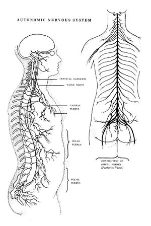
Brain function disruptions occur when NMO affects the brain stem and the hypothalamus which are the main centers that control the autonomic processes in the body. Some of its symptoms include[10]:
- Uncontrollable hiccups
- Pruritus
- Nausea and vomiting
- Diplopia and nystagmus
- Palsy
- Vertigo and ataxia
- Trigeminal neuralgia
Diagnostic Procedures[edit | edit source]
The diagnostic criteria for Neuromyelitis Optica require the presence of history of optic neuritis and transverse myelitis in addition to 3 other supportive criteria which are[11]:
- Magnetic Resonance Imaging (MRI) not diagnostic of Multiple Sclerosis (MS) at disease onset
- Spinal MRI with a contiguous lesion ≥ 3 segments
- Aquaporin-4 immunoglobulin G (AQP4-IgG) seropositivity (Blood Test)
In addition to that other findings which may raise suspicion of NMO include:
- For ON, patients with severe vision loss or visual field depression and MRI findings of posterior optic nerve or chiasm involvement of extensive visual pathway lesions[12].
- For TM, the presence of a longitudinally extensive spinal cord or central cord lesion. In addition to that, cerebrospinal fluid (CSF) findings suggestive of NMO presence include a pleocytosis greater than 50 cells per microliter, a high percentage of polymorphonuclear cells, or the presence of eosinophils[13].
Differential Diagnosis[edit | edit source]
- Multiple sclerosis
- Guillain Barre Syndrome
- Disc herniation
- Transverse myelitis
- Parainfectious myelitis
- Tumour
- Epidural or subdural hematoma
- Epidural and/or paraspinal abscess
Medical Management / Interventions[edit | edit source]
The disease is not curable, however, proper management may ensure the patient live good quality life. Medical intervention may involve; [11]
- Intravenous corticosteroids
- Immunosuppressants
- Plasmapheresis
Physiotherapy Management[edit | edit source]
As stated earlier, no known cure has been found for the disease yet. However, Physiotherapy management is essential for possible future remission as interventions focuses on the clinical presentations which are most times physical disabilities. Physiotherapy interventions may include;
- Gait training
- Wheelchair training
- Pain reduction / management
- Transfers
- Reduce risk of pressure ulcers
- Aid control to spasticity
- Strengthening programme
- Stretching programme
References[edit | edit source]
- ↑ Smith AM, Czyz CN. Neuroanatomy, Cranial Nerve 2 (Optic). InStatPearls. StatPearls Publishing. 2022
- ↑ Kowarik MC, Soltys J. Bennett JL. The Treatment of Neuromyelitis Optica. Journal of Neuro-Ophthalmology. 2014. 34 (1): 70–82
- ↑ 3.0 3.1 3.2 Barkhof F, Koeller KK (February 2020). 13 Demyelinating Diseases of the CNS (Brain and Spine) . In: Hodler J, Kubik-Huch RA, von Schulthess GK (eds.). Diseases of the Brain, Head and Neck, Spine 2020–2023: Diagnostic Imaging . IDKD Springer Series. Cham, Switzerland: Springer. pp. 165–176.
- ↑ Neuromyelitis Optica. Neuromyelitis Optica | Johns Hopkins Medicine. Available at: https://www.hopkinsmedicine.org/health/conditions-and-diseases/neuromyelitis-optica (accessed 17/2/2023)
- ↑ Paul F, Jarius S, Aktas O, Bluthner M, Bauer O, Appelhans H, Franciotta D, Bergamaschi R, Littleton E, Palace J, Seelig HP. Antibody to aquaporin 4 in the diagnosis of neuromyelitis optica. PLoS medicine. 2007 Apr;4(4):e133.
- ↑ Waters P, Jarius S, Littleton E, Leite MI, Jacob S, Gray B, Geraldes R, Vale T, Jacob A, Palace J, Maxwell S. Aquaporin-4 antibodies in neuromyelitis optica and longitudinally extensive transverse myelitis. Archives of neurology. 2008 Jul 14;65(7):913-9.
- ↑ Jarius S, Wildemann B, Paul F. Neuromyelitis optica: clinical features, immunopathogenesis and treatment. Clinical & Experimental Immunology. 2014 May;176(2):149-64.
- ↑ Toosy AT, Mason DF, Miller DH. Optic neuritis. The Lancet Neurology. 2014 Jan 1;13(1):83-99.
- ↑ Frohman EM, Wingerchuk DM. Transverse myelitis. New England Journal of Medicine. 2010 Aug 5;363(6):564-72.
- ↑ Cho EB, Han CE, Seo SW, Chin J, Shin JH, Cho HJ, Seok JM, Kim ST, Kim BJ, Na DL, Lee KH. White matter network disruption and cognitive dysfunction in neuromyelitis optica spectrum disorder. Frontiers in neurology. 2018 Dec 17;9:1104.
- ↑ 11.0 11.1 Wingerchuk DM, Lennon VA, Pittock SJ, Lucchinetti CF, Weinshenker BG. Revised Diagnostic Criteria for Neuromyelitis Optica. Journal of Neurology. 2006. 66 (10): 1485–9.
- ↑ Fernandes DB, Ramos Rde I, Falcochio C, Apostolos-Pereira S, Callegaro D, Monteiro ML. Comparison of visual acuity and automated perimetry findings in patients with neuromyelitis optica or multiple sclerosis after single or multiple attacks of optic neuritis. J Neuroophthalmol. 2012;32:102–106.
- ↑ Jarius S, Paul F, Franciotta D, Ruprecht K, Ringelstein M, Bergamaschi R, Rommer P, Kleiter I, Stich O, Reuss R, Rauer S. Cerebrospinal fluid findings in aquaporin-4 antibody positive neuromyelitis optica: results from 211 lumbar punctures. Journal of the neurological sciences. 2011 Jul 15;306(1-2):82-90.


