Zoonotic Diseases
Original Editors - Kristy Rizzo from Bellarmine University's Pathophysiology of Complex Patient Problems project.
Top Contributors - Kristine Rizzo, Lucinda hampton, Elaine Lonnemann, Admin, Tony Lowe, Kim Jackson, WikiSysop, 127.0.0.1 and Nupur Smit Shah
Definition/Description[edit | edit source]
A zoonotic disease is any disease or infection that is transmissible from vertebrate animals to humans. [1],[2]
Classes of Zoonoses[edit | edit source]
1. Viral Zoonoses[edit | edit source]
Most Common Viral Zoonoses[edit | edit source]
Ehrlichiosis[edit | edit source]
Please see below under Bacterial Zoonoses. Noted here due to diagnostic techniques used.
Rickettsia (Rocky Mountain Spotted Fever)[edit | edit source]
Please see below under Bacterial Zoonoses. Noted here due to diagnostic techniques used.
Rabies[edit | edit source]
A viral disease associated with mammals, including dogs, cats, horses, and wildlife.[3] Rabies can be transmitted through bites, scratches, aerosolized respiratory secretions, and saliva. [1]It may take several weeks or even a few years for people to show symptoms after getting infected with rabies, but usually people start to show signs of the disease 1 to 3 months after the virus infects them. The early signs of rabies can be fever or headache, but this changes quickly to nervous system signs, such as confusion, sleepiness, or agitation. Once someone with rabies infection starts having these symptoms, that person usually does not survive.[3] For this reason, all animal health care workers should be vaccinated against rabies and should have their titers checked every other year.[1] Many kinds of animal can pass rabies to people. Wild animals are much more likely to carry rabies, especially raccoons, skunks, bats, foxes, and coyotes. However, dogs, cats, cattle, or any warm-blooded animal can pass rabies to people.[3]
West Nile Virus[edit | edit source]
A viral disease spread by mosquitoes which can affect birds, horses, and other mammals[3]
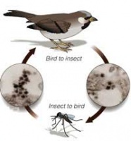
Mosquitoes become infected when they feed on infected birds. Infected mosquitoes can then spread WNV to humans and other animals when they bite. About one in 150 people infected with WNV will develop severe illness. The severe symptoms can include high fever, headache, neck stiffness, stupor, disorientation, coma, tremors, convulsions, muscle weakness, vision loss, numbness and paralysis. These symptoms may last several weeks, and neurological effects may be permanent.
Up to 20 percent of the people who become infected have symptoms such as fever, headache, and body aches, nausea, vomiting, and sometimes swollen lymph glands or a skin rash on the chest, stomach and back.
Symptoms can last for as short as a few days, though even healthy people have become sick for several weeks. Approximately 80 percent of people (about 4 out of 5) who are infected with WNV will not show any symptoms at all. People typically develop symptoms between 3 and 14 days after they are bitten by the infected mosquito. There is no specific treatment for WNV infection. In cases with milder symptoms, symptoms pass on their own. In more severe cases, people usually need to go to the hospital where they can receive supportive treatment including intravenous fluids, help with breathing, and nursing care.[4] Personal protective measures are the primary way to avoid contracting the virus.[1]
Equine Encephalitis
[edit | edit source]
A mosquito borne infection normally maintained in nature by a cycle from an arthropod vector to a vertebrate reservoir host.[1] Although some people experience it only as a mild illness, eastern equine encephalitis is fatal in about one-third of the cases. Symptoms of eastern equine encephalitis usually appear three to 10 days after a bite by an infected mosquito.[5] A vaccine exists for horses but not for humans.[1] Personal protective measures are the primary way to avoid contracting the virus.[1]
A. Diagnostic Tests/Lab Tests/Lab Values [edit | edit source]
1. ELISA (Enzyme Lined Immuno-Sorbent Assay)- detects the actual disease. May not be sensitive enough to detect infection in the asymptomatic carrier.[1]
2. PCR (Polymerase Chain Reaction)- amplifies minute quantites of microbial DNA or RNA allowing for recognition of latent phases of viral diseases. May yield false positive result for the disease but give a true positive for infection within the agent.[1]
3. Serology (Indirect Fluorescent Antibody Testing)- used to detect Rickettsial organisms which cause Ehrlichiosis and Rickettsia[1]
2. Bacterial Zoonoses[edit | edit source]
Most Common Bacterial Zoonoses[edit | edit source]
Anthrax[edit | edit source]
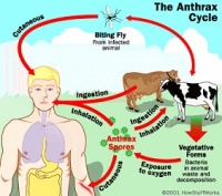
Anthrax is an acute infectious disease caused by the spore-forming bacterium Bacillus anthracis[6], a microbe that lives in the soil.[7] Anthrax most commonly occurs in wild and domestic mammals (cattle, sheep, goats, camels, antelopes, and other herbivores), but it can also occur in humans when they are exposed to infected animals or to tissue from infected animals.[6] Anthrax can occur in three forms: cutaneous, inhalation, and gastrointestinal.[6][7]
Bartonella (Cat Scratch Fever) [edit | edit source]
Cat scratch disease (CSD) is a bacterial disease caused by Bartonella henselae. Most people with CSD have been bitten or scratched by a cat and developed a mild infection at the point of injury. Lymph nodes, especially those around the head,
neck, and upper limbs, become swollen. Additionally, a person with CSD may experience fever, headache, fatigue, and a poor appetite.[3] Bartonella will begin in the human as a pustule that will gradually progress to regional lymphadenopathy which can last for months (or become a systemic illness in immunocompromised patients). In addition to the cat or dog scratch, bartonella can also be transmitted through the feces of fleas.[1]
Borrelia (Lyme Disease)[edit | edit source]
Lyme disease is a bacterial disease caused by Borrelia burgdorferi. People get lyme disease
when they are bitten by ticks carrying this bacteria. Within 1 to 2 weeks of being infected, people may have a "bull's-eye" rash with fever, headache, and muscle or joint pain.
Some people have Lyme disease and do not have any early symptoms. Other people have a fever and other "flu-like" symptoms without a rash. After several days or weeks, the bacteria may spread throughout the body of an infected person. These people can get symptoms such as rashes in other parts of the body, pain that seems to move from joint to joint, and signs of inflammation of the heart or nerves. If the disease is not treated, a few patients can get additional symptoms, such as swelling and pain in major joints or mental changes, months after getting infected.[3] Additional symptoms in humans include encephalomyelitis, meningitis, radiculo-neuropathy, and cranial neuropathies (most notably facial paralysis).[1]
Clinical signs in the canine patient may include acute lameness due to polyarthritis.[1]
Brucellosis[edit | edit source]
Brucellosis is a zoonotic infection transmitted from animals to humans by the ingestion of infected food
products, direct contact with an infected animal, or inhalation of aerosols.[8] The timely and accurate diagnosis of human brucellosis continues to challenge clinicians because of its non-specific clinical features, slow growth rate in the blood culture, and the complexity of its serodiagnosis.[8] Four different species of Brucella are known to infect humans: B. abortus (cattle), B. suis (swine), B. melitensis (goats/sheep) and B. canis (dogs). B. abortus and B. canis cause a mild fever whereas B. suis causes a more severe infection which can lead to destruction of the lymphoreticular organs and kidney. B.melitensis is the cause of most severe prolonged recurring disease. The bacteria enter the human host through mucous membranes. The bacteria localize in the regional lymph nodes, where they proliferate intracellularly. If the Brucella organisms are not destroyed or contained in the lymph nodes, the bacteria are released from the lymph nodes resulting in septicemia.[9]
Brucellosis in human beings is rarely fatal, but can lead to severe debilitation and disability[8] Symptoms include fever, chills, sweats, fatigue, myalgia, profound muscle weakness, and anorexia.[9] Diagnostic methods for brucellosis are primarily based on serology, with the LPS smooth chains producing the greatest immunological responses in various hosts.[8],[9] There is currently no human vaccine available.[10]
Dogs infected with brucellosis may be asymptomatic or may show systemic disease such as discospondylitis. Back pain or paresis may be differential diagnoses for these patients.[1]
Ehrlichia[edit | edit source]
Ehrlichiosis is the general name used to describe several bacterial diseases that affect animals and humans. Human ehrlichiosis is a disease caused by at least three different ehrlichial species in the United States: Ehrlichia chaffeensis, Ehrlichia ewingii, and a third Ehrlichia species provisionally called Ehrlichia muris-like (EML). Ehrlichiae are transmitted to humans by the bite of an infected tick. Symptoms usually occur within 1-2 weeks following a tick bite[11][12] and include fever, headache, fatigue, and muscle aches. Ehrlichios is is diagnosed based on symptoms, clinical presentation, and later confirmed with specialized laboratory tests. Treatment for adults and children of all ages is doxycycline.[11] Same diagnositc techniques are used as with viruses[1]
Leptospirosis[edit | edit source]
is caused by many different species of the spirochete Leptospira.[1][13] Dogs can be a vector for human disease, and humans are infected by exposure to urine, blood, or tissue from an infected dog. If shed in urine, the organism can survive in an environment between 0°-25°C. Prevention can be improved by good sanitation practices and vaccination of dogs. The most common means of transmission is through water contact with skin wounds or mucous membranes. Leptospires invade organs including the kidneys, liver[1][13], spleen, central nervous system[1][13], eyes, and genital tract. Any dogs which are suspected of shedding leptospira should be treated with doxycycline.[1]
Plague (Yersinia enterocolitica, Yersinia pestis) [edit | edit source]
A rare bacterial disease associated with wild rodents, cats, and fleas.[3]Yersiniosis is a disease caused by the bacterium Yersinia enterocolitica[14], or yersinia pestis (a gram negative coccobacillus).[15] Plague (yersinia pestis) is the disease known in the middle ages as black death due to the gangrene and blackening of various body parts associated with this disease.[9] However people with yersiniosis can have different symptoms depending on age.

Symptoms can begin 4 to 7 days after infection. Young children usually have fever, stomach pain, and diarrhea. Adults will sometimes feel pain on their right side and may have a fever. Usually, these signs go away after about 3 weeks but sometimes pain in joints, such as knees or wrists, can start after that and last for several months.[14] Another symptom associated with yersinia pestis is swollen lymph nodes. The infection spreads into the lymph nodes causing swelling, hot temperature, and tenderness.[9]
Animals can pass Yersinia enterocolitica in their stool, and people can get sick from contact with infected feces. Several kinds of animals can carry this disease such as cats, dogs, horses, cows, rodents, and rabbits, and pigs.[14] The pulmonary form is spread by airborne or droplet infection. Human infections from nonrodent species or carrier fleas[9] usually result from direct contact with infected tissues, by scratch or bite injuries, and handling of infected animals.[15]
Rickettsia (Rocky Mountain Spotted Fever)[edit | edit source]
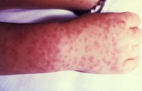
A disease caused by the bacterium Rickettsia rickettsii, which is carried by ticks. People usually start having fevers and feeling nauseous about a week after being bitten by a tick. A few days after the fever begins people will often have a rash, usually on the arms or ankles. They also may have pain in their joints, stomach pain, and diarrhea.[3]
People get this disease when they are bitten by a tick that is carrying the bacterium R. rickettsia. Because ticks on dogs can be infected with R. rickettsii, dogs and people can get Rocky Mountain spotted fever from the same ticks. These ticks can also bite other animals and pass Rocky Mountain spotted fever to them. When you remove ticks from any animal, the crushed tick or its parts can also pass this disease through any cuts or scrapes on your skin.[3]
Diagnositc techniques used are the same as with viruses.[3]
Methicillin Resistant Staphylococcus Aureus (MRSA)[edit | edit source]
The most common MRSA infections between pets and humans are skin, soft-tissue, and surgical infections. Dog or cat bites can result in infection, caused by bacteria from the animal's mouth and on the patients' body. Animals are potential reservoirs of MSRA infection due to increasing prevalence of community-acquired MRSA (CA-MRSA) in humans and domestic animals such as dogs, cats and horses. MRSA-associated infections in pets are typically acquired from their owners and can potentially cycle between pets and humans.[2]
Treatment of MRSA infections in pets is similar to that used in humans. Resistant to penicillin and methicillin, CA-MRSA infections can still be treated with other common-use antibiotics. CA-MRSA most often enters the body through a cut or scrape and appears in the form of a skin or soft tissue infection, such as a boil or abscess. The involved site is red, swollen, and painful and is often mistaken for a spider bite. Though rare, CA-MRSA can develop into more serious invasive infections, such as bloodstream infections or pneumonia, leading to a variety of other symptoms including shortness of breath, fever, chills, and death. CA-MRSA can be particularly dangerous in children because their immune systems are not fully developed.[2]
Streptococcus suis[edit | edit source]
Streptococcus suis is a zoonotic pathogen that infects pigs and can occasionally cause serious infections in humans. S. suis infections occur sporadically in humans throughout Europe and North America, but a recent major outbreak has been described in China with high levels of mortality. The mechanisms of S. suis pathogenesis in humans and pigs are poorly understood.[16]
A. Diagnostic Tests/Lab Tests/Lab Values[edit | edit source]
1. Blood Culture is the gold standard in the diagnosis of bacterial infections including brucellosis[8]
3. Fungal Zoonoses[edit | edit source]
Most Common Fungal Zoonoses[edit | edit source]
Dermatomycoses[edit | edit source]
Dermatomycoses are caused by fungal spores which remain viable for long periods on carrier animals. Exposure to reservoir hosts harboring different dermatophytes determines the type and incidence of infection in humans. Pets may also acquire disease from humans. Dermatomycoses can be transmitted directly or indirectly through contact with asymptomatic animals or skin lesions on infected animials, contaminated bedding or equipment, fungi in the air, in dust, or on surfaces of the room. The disease in rodents or in cats is often asymptomatic and not recognized until people are affected, but dogs will often show classic skin lesions. Varying severity of dermatitis occurs with local loss of hair. Deeper invasion produces a mild inflammatory reaction which increases in severity with the development of hypersensitivity. In humans, the disease is often mild and self limiting. Scaling, redness, and occasionally vesicles or fissures will be present with thickening and discoloring of nails. A circular lesion with a clear center may also be present.[17]
Systemic Fungal Diseases (indirect zoonoses)[edit | edit source]
Blastomycosis[edit | edit source]
Blastomycosis generally results from inhalation of Blastomyces dermatitidis conidia following exposure to contaminated soil in an endemic area. Primary infections commonly involve the lungs, although secondary dissemination to other body sites may occur.[18]
Coccidioidomycosis[edit | edit source]
Coccidioidomycosis is an infection with the spores of the fungus Coccidioides immitis. It is most commonly seen in the
desert regions of the southwestern United States and in Central and South America. The infection starts in the lungs and is contracted by breathing in fungal particles from soil. Most people with this infection do not have symptoms. Some may have cold or flu-like symptoms or symptoms of pneumonia. If symptoms occur, they typically start 5 to 21 days after being
exposed to the fungus. Symptoms include, change in mental status, chest pain, cough (possibly hemoptysis, fever, headache, joint
stiffness/pain, loss of appetite, muscle aches, neck stiffness, night sweats, painful red lumps on lower legs (erythema nodosum), sensitivity to light, weight loss, and wheezing.[19]
Histoplasmosis[edit | edit source]
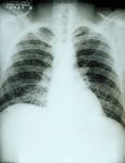
Histoplasmosis is a fungal disease associated with bat guano (stool).[3] The infection enters the body through the lungs. Histoplasma fungus grows as a mold in the soil, and infection results from breathing in airborne particles. Soil contaminated with bird or bat droppings may have a higher concentration of histoplasma. There may be a short period of active infection, or it can become chronic and spread throughout the body. Histoplasmosis may have no symptoms. Most people who do develop symptoms will have a flu-like syndrome and lung (pulmonary) complaints related to pneumonia or other lung involvement. Those with chronic lung disease (such as emphysema and bronchiectasis) are at higher risk of a more severe infection.[20]
Cryptococcosis[edit | edit source]
Cryptococcosis is a fungal disease caused by Cryptococcus neoformans or Cryptococcus gattii. This fungus is found in the droppings of wild birds (such as pigeons). When dried bird droppings are stirred up, this can transfer dust containing Cryptococcus neoformans into the air. People can stir up this dust and then breathe it in when they work, play, or walk in areas where birds have been. Pets, such as dogs and cats, can also get sick with cryptococcosis from this dust, but people do not get cryptococcosis from directly from dogs and cats. Cryptococcosis can cause serious symptoms of lung, brain and spinal cord disease, such as headaches, fever, cough, shortness of breath, and night sweats.[21]
Diagnostic Tests/Lab Tests/Lab Values[edit | edit source]
Diagnostic tests used include:
Sputum culture and examination[22]
Tissue biopsy[22]
Urine culture[22]
Blood, urine, or sputum tests to look for signs of histoplasmosis infection[20]
Spinal tap to look for signs of infection in cerbrospinal fluid (CSF)[20]
4. Parasitic Zoonoses
[edit | edit source]
Most Common Parasitic Zoonoses[edit | edit source]
Toxocara Canis (Roundworm)[edit | edit source]
Toxocara canis is a parasitic disease associated with cats, dogs and their environment.[3] Toxocara canis is an infection transmitted from animals to humans caused by the parasitic roundworms commonly found in the intestine of dogs (Toxocara canis) and cats (T. cati). The most common Toxocara parasite of concern to humans is T. canis, which puppies usually contract from the mother before birth or from her milk. The larvae mature rapidly in the puppy’s intestine; when the pup is 3 or 4 weeks old, they begin to produce large numbers of eggs that contaminate the environment through the animal’s stool. The eggs soon develop into infective larvae. Humans can become infected after accidentally ingesting infective Toxocara eggs in soil or other contaminated surfaces. In most cases, Toxocara infections are not serious, and many people, especially adults infected by a small number of larvae, may not notice any symptoms. The most severe cases are rare, but are more likely to occur in young children, who often play in dirt, or eat dirt (pica) contaminated by dog or cat stool.[23]
Ancylostoma Caninum (hookworm)[edit | edit source]
Ancylostoma caninum is a parasitic disease associated with dogs and their environment.[3] Animals can indirectly pass hookworm to humans. Animals that are infected pass hookworm eggs in their stools. The eggs can hatch into larvae, and both eggs and larvae may be found in dirt where animals have been. Eggs or larvae can get into the human body through direct contact with the dirt. For example, this can happen if a child is walking barefoot or playing in an area where dogs or cats have been (especially puppies or kittens). If a person accidentally contracts animal hookworm eggs, then the larvae that hatch out of the eggs can reach the intestine and cause bleeding, inflammation (swelling), and abdominal pain. People can get painful and itchy skin infections when animal hookworm larvae move through their skin.[24]
Echinococcosis (tapeworm)[edit | edit source]
The adult worm affects various final hosts such as domestic dogs and wild carnivores like foxes, coyotes, wolves and jackals. The eggs are spread through their faeces. Intermediate hosts are sheep, cattle, swine, goats,
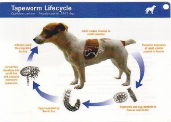
equines, camelids and cervids. They acquire the infection by ingestion of the infective eggs during grazing. Carnivores acquire the infection by ingestion of infected raw material of the intermediate hosts (mostly viscera). Like the intermediate hosts, humans acquire the infection by ingestion of infective eggs.
Echinococcosis is mainly maintained in the dog-sheep-dog cycle. The infection is transmitted to dogs when they are feed infected viscera of sheep during the home-slaughter of sheep. The eggs are found on the surface of faecal matter of dogs, and they can accumulate in the perianal region of dogs. The dog carries the eggs on it's tongue and snout to different parts of its body. Direct contact with dogs is an important mode of transmission to humans, as is consumption of vegetables and water contaminated with infected dog faeces. Humans are accidental intermediate hosts and are not able to transmit the disease.[25]
Ectoparasites (ticks and fleas)
[edit | edit source]
Rocky Mountain Spotted Fever (see above)[edit | edit source]
Ehrlichiosis (see above)[edit | edit source]
Plague (see above)[edit | edit source]
Lyme Disease (see above)[edit | edit source]
Diagnostic Tests/Lab Tests/Lab Values[edit | edit source]
Diagnostic tests used include:
Complete Blood Count (CBC)[26][27][28]
Stool ova and parasites exam[26]
Physical examination[28]
Prevention[edit | edit source]
The best way to protect oneself from many of these zoonotic diseases is to practice good hygiene after handling animals or their waste. Washing hands thoroughly with hot, soapy water after any contact will help prevention contraction of zoonotic diseases.[2] In addition screening newly received animals, conducting a routine sanitization of the contaminated environment, equipment, and caging, wearing gloves and protective clothing will help decrease the possiblity of contracting a zoonotic disease.[17]
The four principal means of preventing spread of zoonoses are[1]
1. parasite recognition and control
2. vaccination programs
3. sanitation methods
4. behavior training to prevent bites and scratches
Differential Diagnoses[edit | edit source]
For the animal: lameness, weakness, cranial cruciate ligament rupture, hypoadrenocorticism (Addison's Disease)[29]
Resources[edit | edit source]
For more information on rabies vaccines, visit http://www.cdc.gov/vaccines/pubs/vis/downloads/vis-rabies.pdf
For biosecurity and infection control guidelines for vets, visit http://csuvets.colostate.edu/biosecurity/
Recent Related Research (from Pubmed)[edit | edit source]
Recent Related Research (from Pubmed)
Failed to load RSS feed from http://eutils.ncbi.nlm.nih.gov/entrez/eutils/erss.cgi?rss_guid=14a2MZa9gZvcIZpSDTFlxN1kXWpZ0UuViHP7shbdeYBeIWkgUM: Error parsing XML for RSS
References[edit | edit source]
see adding references tutorial.
- ↑ 1.00 1.01 1.02 1.03 1.04 1.05 1.06 1.07 1.08 1.09 1.10 1.11 1.12 1.13 1.14 1.15 1.16 1.17 1.18 1.19 Van Dyke JB. Veterinary zoonoses, what you need to know before you treat that puppy! American Physical Therapy Association Combined Sections Meeting; 2011 Feb 11; New Orleans, Louisianna.
- ↑ 2.0 2.1 2.2 2.3 Oregon Veterinary Medical Association. Zoonotic Diseases and Horses.fckLRhttp://oregonvma.org/care-health/zoonotic-diseases-horses. (accessed 8 March 2011).
- ↑ 3.00 3.01 3.02 3.03 3.04 3.05 3.06 3.07 3.08 3.09 3.10 3.11 3.12 Centers for Disease Control and Prevention. Healthy Pets Healthy People. http://www.cdc.gov/healthypets/browse_by_diseases.htm (accessed 26 Feb 2011).
- ↑ Centers for Disease Control and Prevention. Division of Vector-Borne Infectious Diseases. West-Nile Virus. http://www.cdc.gov/ncidod/dvbid/westnile/wnv_factsheet.htm (accessed 27 Feb 2011).
- ↑ Mayo Foundation for Medical Education and Research. Encephalitis. http://www.mayoclinic.com/health/encephalitis/DS00226/DSECTION=causes (accessed 27 Feb 2011).
- ↑ 6.0 6.1 6.2 Centers for Disease Control and Prevention. Emergency Preparedness and Response. Questions and Answers about Anthrax. http://www.bt.cdc.gov/agent/anthrax/faq/ (accessed 2 March 2011)
- ↑ 7.0 7.1 U.S. National Library of Medicine. National Institute of Health. Medline Plus. Anthrax.http://www.nlm.nih.gov/medlineplus/anthrax.html (accessed 2 March 2011).
- ↑ 8.0 8.1 8.2 8.3 8.4 Christopher S, Umapathy BL, Ravikumar KL. Brucellosis: Review on the recent trends in pathogenicity and laboratory diagnosis. J Lab Physicians 2010;2:55-60
- ↑ 9.0 9.1 9.2 9.3 9.4 9.5 University of South Carolina School of Medicine. Microbiology and Immunology On-line. Bacteriology Chapter Seventeen. Zoonoses. http://pathmicro.med.sc.edu/ghaffar/zoonoses.htm (accessed 10 March 2011).
- ↑ Teske SS, Huang Y, Tamrakar SB, Bartrand TA, Weir MH, Haas CN. Animal and human dose-response models for Brucella species. Risk Anal 2011; http://www.ncbi.nlm.nih.gov/pubmed/21449960?dopt=Abstract (accessed 4 April 2011).
- ↑ 11.0 11.1 Centers for Disease Control and Prevention. Ehrlichiosis. http://www.cdc.gov/ehrlichiosis/ (accessed 27 Feb 2011).
- ↑ Mayo Foundation for Medical Education and Research. Ehrlichiosis. http://www.mayoclinic.com/health/ehrlichiosis/DS00702 (accessed 27 Feb 2011).
- ↑ 13.0 13.1 13.2 Centers for Disease Control and Prevention. Leptospirosis. http://www.cdc.gov/leptospirosis/ (accessed 3 March 2011).
- ↑ 14.0 14.1 14.2 Centers for Disease Control and Prevention. Yersinia enterocolitica and Pigs. http://www.cdc.gov/healthypets/diseases/yersinia.htm (accessed 8 March 2011).
- ↑ 15.0 15.1 Iowa State University. Institutional Biosafety Committee. Guidance & Education. Zoonotic Disease Fact Sheets: yersinia pestis. http://compliance.iastate.edu/ibc/guide/zoonoticfactsheets/Yersinia%20Pestis.pdf (accessed 8 March 2011).
- ↑ Holden et al. Rapid evolution of virulence and drug resistance in the emerging zoonotic pathogen streptococcus suis. PLoS One. 2009; 4(7). e6072. http://www.ncbi.nlm.nih.gov/pmc/articles/PMC2705793/(accessed 8 March 2011).
- ↑ 17.0 17.1 Iowa State University. Institutional Biosafety Committee. Guidance & Education. Zoonotic Disease Fact Sheets: dermatomycoses. http://compliance.iastate.edu/ibc/guide/zoonoticfactsheets/Dermatomycoses.pdf (accessed 8 March 2011).
- ↑ Veligandla et al. Delayed diagnosis of osseous blastomycosis in two patients following environmental exposure in nonendemic areas. Am. J. Clin. Pathol. 118 (4): 536–41. http://ajcp.ascpjournals.org/content/118/4/536 (accessed 8 March 2011).
- ↑ National Center for Biotechnology Information, U.S. National Library of Medicine. Coccidioidomycosis. http://www.ncbi.nlm.nih.gov/pubmedhealth/PMH0002299/ (accessed 8 March 2011).
- ↑ 20.0 20.1 20.2 20.3 20.4 20.5 National Center for Biotechnology Information, U.S. National Library of Medicine. Histoplasmosis. http://www.ncbi.nlm.nih.gov/pubmedhealth/PMH0002073/ (accessed 8 March 2011).
- ↑ Centers for Disease Control and Prevention. Healthy Pets Healthy People. Cryptococcosis. http://www.cdc.gov/healthypets/diseases/crptococcus.htm (accessed 8 March 2011).
- ↑ 22.0 22.1 22.2 22.3 22.4 22.5 National Center for Biotechnology Information, U.S. National Library of Medicine. Blastomycosis. http://www.ncbi.nlm.nih.gov/pubmedhealth/PMH0001163/ (accessed 4 April 2011).
- ↑ Centers for Disease Control and Prevention. Parasites. Toxocariasis. http://www.cdc.gov/parasites/toxocariasis/gen_info/faqs.html (accessed 8 March 2011).
- ↑ Centers for Disease Control and Prevention. Healthy Pets Healthy People. Hookworm Infection and Animals. http://www.cdc.gov/healthypets/diseases/hookworm.htm (accessed 8 March 2011).
- ↑ World Health Organization. Zoonoses and Veterinary Public Health. Cystic echinococcosis and multilocular echinococcosis. http://www.stanford.edu/group/parasites/ParaSites2006/Echinococcus/index.html (accessed 8 March 2011).
- ↑ 26.0 26.1 National Center for Biotechnology Information, U.S. National Library of Medicine. Hookworm. http://www.ncbi.nlm.nih.gov/pubmedhealth/PMH0001653/ (accessed 4 April 2011).
- ↑ National Center for Biotechnology Information, U.S. National Library of Medicine. Visceral larva migrans. http://www.ncbi.nlm.nih.gov/pubmedhealth/PMH0001657/ (accessed 4 April 2011).
- ↑ 28.0 28.1 National Center for Biotechnology Information, U.S. National Library of Medicine. Echinococcus. http://www.ncbi.nlm.nih.gov/pubmedhealth/PMH0001697/ (accessed 4 April 2011).
- ↑ Van Dyke JB. Veterinary red flags, endocrine, metabolic and medical syndromes that may be lurking in your canine rehabilitation patient. American Physical Therapy Association Combined Sections Meeting; 2011 Feb 11; New Orleans, Louisianna.


