Thoracic Anatomy
This article or area is currently under construction and may only be partially complete. Please come back soon to see the finished work! (12/04/2020)
Original Editor - Your name will be added here if you created the original content for this page.
Top Contributors - Lucinda hampton and Kim Jackson
Introduction[edit | edit source]
This guide gives a general overview of the anatomy of the thoracic spine. The sagittal plane alignment of the Thoracic spine is on average 35% (normal range is 20° to 50°).[1]
Important Structures
The important parts of the thoracic spine include
- bones
- joints
- structure/motion
- nerves
- connective tissues
- muscles
- spinal segment[2]
Bones[edit | edit source]
The human spine is made up of 24 spinal bones, called vertebrae. Vertebrae are stacked on top of one another to create the spinal column. The spinal column is the body’s main upright support.
- The middle 12 Thoracic vertebrae make up the thoracic spine.
- T5-T8 tend to be the most “typical” in that they contain features present in all thoracic vertebrae. T5-T8 have the greatest rotation ability of the thoracic region.
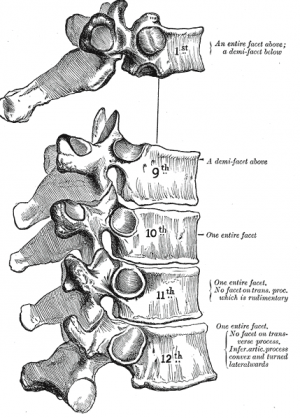
- T1 - superior costal facets are “whole” costal facets. They alone articulate with the first rib; C7 has no costal facets. T1 does, however, have typical inferior demifacets for articulation with the second rib. Vertebral prominens name given to long prominent spinous process found at T1
- T11 and T12 are atypical - contain a single pair, “whole,” costal facet that articulate with the 11 and 12 ribs, respectively. They also lack facets on the transverse processes.[3]
- The primary characteristic of the thoracic vertebrae is the presence of costal facets. There are 6 facets per thoracic vertebrae: 2 on the transverse processes and 4 demifacets. The facets of the transverse processes articulate with the tubercle of the associated rib. The demifacets are bilaterally paired and located on the superior and inferior posterolateral aspects of the vertebrae. They are positioned so that the superior demifacet of inferior vertebrae articulates with the head of the same rib that articulates with the inferior demifacet of a superior rib. For example, the inferior demifacets of T4 and the superior demifacets of T5 articulate with the head of rib 5.
- The length of the transverse processes decreases as the column descends.
- The positioning of the ribs and spinous processes greatly limits flexion and extension of the thoracic vertebrae.
- Thoracic vertebrae have superior articular facets that face in a posterolateral direction.
- The spinous process is long, relative to other regions, and is directed posteroinferiorly. This projection gradually increases as the column descends before decreasing rapidly from T9-T12.[3]
- The intervertebral disc height, is, on average, the least of the vertebral regions.
- Vertebral body size: increases progressively from T1 to T12
- Spinal canal dimensions: varies from T1 to T12[1]
Joints[edit | edit source]
There are 3 types of joints present in the thoracic spine:
Between vertebral bodies – adjacent vertebral bodies are joined by intervertebral discs, made of fibrocartilage. This is a type of cartilaginous joint, known as a symphysis.
Between vertebral arches ie facet joints – formed by the articulation of superior and inferior articular processes from adjacent vertebrae. It is a synovial type joint. In the thoracic spine facet joints angled at:
- 60° to the transverse plane
- 20° to the frontal plane, with the superior facets facing posterior and a little up and laterally and the inferior facets facing anteriorly, down, and medially.
- motion: lateral flexion and rotation; no flexion/extension
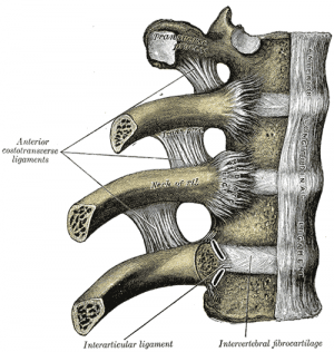
Costovertebral joints, unique to the thoracic spine - consists of the head of the rib articulating with:
- Superior costal facet of the corresponding vertebra
- Inferior costal facet of the superior vertebra
- Intervertebral disc separating the two vertebrae
Within this joint, the intra-articular ligament of head of rib attaches the rib head to the intervertebral disc. Only slight gliding movements can occur at these joints, due to the close articulation of their components.[4]
Structure and Function[edit | edit source]
The anatomy of the Thoracic spine is related directly to its function. The facets and demifacets devoted to rib articulation demonstrate the main function of the thoracic spine.
- The zygapophyseal joints are tight enough to protect vital organs but loose enough to allow for respiratory movements as well as to allow the thoracic segment to have the greatest freedom of rotation of the entire spine.
- The zygapophyseal joints, as well as the relatively thin intervertebral discs, cause the thoracic region to have the least flexion/extension ability of the spine. Prevents downward flexion on heart and lungs
- The increase in vertebral body size as the spinal column descends is directly related to the increased weight-bearing requirement; further down the column, the greater the proportion of body mass that rests upon it.
Sub Heading 3[edit | edit source]
Resources[edit | edit source]
- bulleted list
- x
or
- numbered list
- x
References[edit | edit source]
- ↑ 1.0 1.1 Othrobullets Thoracic spine Anatomy Available from:https://www.orthobullets.com/spine/2070/thoracic-spine-anatomy (last accessed 12.4.2020)
- ↑ eOrthopod Thoracic spine anatomy Available from:https://eorthopod.com/thoracic-spine-anatomy/ (last accessed 12.4.2020)
- ↑ 3.0 3.1 Waxenbaum JA, Futterman B. Anatomy, Back, Thoracic Vertebrae.Available from:https://www.statpearls.com/kb/viewarticle/32138 (last accessed 12.4.2020)
- ↑ Teach me anatomy The Thoracic Spine Available from:https://teachmeanatomy.info/thorax/bones/thoracic-spine/ (last accessed 12.4.2020)


