Takotsubo Cardiomyopathy
Original Editors - Megan Kaiser & Briana Ulanowski from Bellarmine University's Pathophysiology of Complex Patient Problems project.
Top Contributors - Megan Kaiser, Briana Ulanowski, Kim Jackson, Lucinda hampton, Elaine Lonnemann, Wendy Walker, WikiSysop, 127.0.0.1 and Adam Vallely Farrell
Definition/Description[edit | edit source]
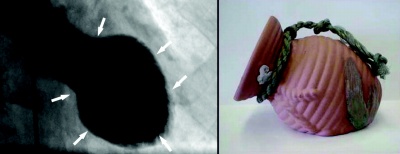
Takotsubo cardiomyopathy occurs when there is an abnormal contraction of the transient left ventricle, creating a balloon shape appearance initially during systole. The Japanese first described the heart condition around 1991. The shape of the heart resembles a Japanese octopus pot with a rounded bottom and narrow neck; hence the name tako-tsubo.[1]
Typically an intense emotional stress can trigger this type of event. High levels of catecholamines are present in these patients and can cause the heart to be stunned temporarily. This is a reversible cardiomyopathy and clinically presents as a myocardial infarction. Individuals that experience this may or may not have a cardiovascular disease.
Other names include:
—Broken heart syndrome
—Ampulla cardiomyopathy
—Stress cardiomyopathy
—Apical ballooning syndrome (ABS)
—Acute left ventricular ballooning[1]
Prevalence[edit | edit source]
Takotsubo cardiomyopathy is a very rare condition and has to be differentiated from myocardial infarct. The incidence of takotsubo cardiomyopathy is 1-2% in patients diagnosed with MI. These individuals are usually postmenopausal females (90%). The average age is 62-75 years old. There are approximately 7,000-14,000 cases of takotsubo cardiomyopathy in the U.S.[2]
Characteristics/Clinical Presentation
[edit | edit source]
Usually takotsubo cadiomyopathy is triggered by emotional stress, physical stress, non-cardiac surgery or procedure (refer to table). The onset is insidious and presents with chest pain at rest.
| Table I. Stressors reported to trigger ABS[3] Emotional stress Death or severe illness or injury of a family member, friend, or pet Receiving bad news—diagnosis of a major illness, daughter's divorce, spouse leaving for war Severe argument Public speaking Involvement with legal proceedings Financial loss—business, gambling Car accident Surprise party Move to a new residence Physical stress Non-cardiac surgery or procedure—cholecystectomy, hysterectomy Severe illness—asthma or chronic obstructive airway exacerbation, connective tissue disorders, acute cholecystitis, pseudomembranous colitis Severe pain—fracture, renal colic, pneumothorax, pulmonary embolism Recovering from general anesthesia Cocaine use Opiate withdrawal Stress test—dobutamine stress echo, exercise sestamibi Thyrotoxicosis |
Signs and Symptoms of Takotsubo include[4]:[5]
• Chest pain
• Dyspnea
• Hypotension
• Syncope
• Mild to moderate HF
• Pulmonary edema
• Lab Changes
• Modest increase in troponin T levels
• Increase in creatine phosphokinase (CPK)
• Elevation in pBNP
• High levels of serum catecholamines
The most common signs and symptoms are chest pain and dyspnea; resembling a myocardial infarction.
Associated Co-morbidities[edit | edit source]
Research has not shown any comorbidities linked with TC.
Medications[edit | edit source]
add text here
Diagnostic Tests/Lab Tests/Lab Values[edit | edit source]
Diagnostic tests used for TC include lab values, echocardiography, cardiac angiography, and electrocardiography.
Lab Values
Troponin T level:
- Mean value of healthy individuals = 0.49 ng/mL,
- Mean peak value of patients with TC = 0.64 ng/mL
Troponin I level:
- Mean value of healthy individuals = 4.2 ng/mL
- Mean peak value of patients with TC = 8.6 ng/mL
Brain natriuretic peptide level is elevated
Catecholamines are elevated in the acute phase
Echocardiography
An echocardiography is used when diagnosing wall-motion abnormalities. The heart will show hypokinesis or akinesis of the middle and apical segment of the left ventricle if the diagnosis is TC.
Cardiac Angiography
The test will show normal coronary arteries in patients with TC.
Electrocardiography (EKG) Changes[6]
Generally, patients could have 4 different EKG changes:
- ST-segment elevation
- T-wave inversion
- Abnormal Q waves
- QT segment prolongation
In a study conducted by Mitsuma et al, they showed that EKG changes could occur in phases:
- ST-segment elevation (immediately)
- T-wave inversion (days 1-3)
- Improvement in T-wave inversion (days 2-6)
- Deeper T-wave inversion
Diagnostic Criteria[7]
Must have all 4 criteria to be diagnosed with ABS
• Transient hypokinesis, akinesis, or dyskinesis of the LV midsegments
• Absence of obstructive coronary artery or angiographic evidence of acute plaque rupture
• New EKG abnormalities or elevated cardiac troponin
• Absence of recent head trauma, intracranial bleeding, pheochromocytoma, myocarditis, and hypertrophic cardiomyopathy
Etiology/Causes[edit | edit source]
add text here
Systemic Involvement[edit | edit source]
There are a number of possible complications in patients who present with takotsubo cardiomyopathy. Left-sided congestive heart failure with pulmonary edema, cardiogenic shock, ventricular fibrillation, left ventricular thrombus formation, and left ventricular free wall rupture are all possibilities. Death is included in this list; however, the mortality rate is only 0-8%, and generally less than 2%.[8]
Medical Management (current best evidence)[edit | edit source]
add text here
Physical Therapy Management (current best evidence)[edit | edit source]
add text here
Alternative/Holistic Management (current best evidence)[edit | edit source]
add text here
Differential Diagnosis[edit | edit source]
add text here
Case Reports/ Case Studies[edit | edit source]
Resources
[edit | edit source]
add appropriate resources here
Recent Related Research (from Pubmed)[edit | edit source]
see tutorial on Adding PubMed Feed
Extension:RSS -- Error: Not a valid URL: addfeedhere|charset=UTF-8|short|max=10
References[edit | edit source]
see adding references tutorial.
- ↑ 1.0 1.1 Nussinovitch U, Goitein O, Nussinovitch N. Distinguishing a Heart Attack From the “Broken Heart Syndrome” (Takotsubo Cardiomyopathy). Journal of Cardiovascular Nursing. 2011; 1-6.
- ↑ Prasad A. Lerman A, Rihal CS. Apical ballooning syndrome (Tako-tsubo or stress cardiomyopathy): A mimic of acute myocardial infarction. American Heart Journal. 2008; 155 (3):408-417.
- ↑ Prasad A. Lerman A, Rihal CS. Apical ballooning syndrome (Tako-tsubo or stress cardiomyopathy): A mimic of acute myocardial infarction. American Heart Journal. 2008; 155(3):408-417.
- ↑ Coons JC, Barnes M, Kusick K. Takotsubo cardiomyopathy. Am J Health-Syst Pharm. 2009;66:562-66.
- ↑ Prasad A. Lerman A, Rihal CS. Apical ballooning syndrome (Tako-tsubo or stress cardiomyopathy): A mimic of acute myocardial infarction. American Heart Journal. 2008;155(3):408-417.
- ↑ Coons JC, Barnes M, Kusick K. Takotsubo cardiomyopathy. Am J Health-Syst Pharm. 2009;66:562-66.
- ↑ Coons JC, Barnes M, Kusick K. Takotsubo cardiomyopathy. Am J Health-Syst Pharm. 2009;66:562-66.
- ↑ Barker S, Solomon H, Bergin JD. Electrocardiographic ST-segment elevation: Takotsubo cardiomyopathy versus ST-segment elevation myocardial infarction – A case series. Amer J of Emer Med 2009; 27: 220-226.


