TMJ Anatomy
Original Editor - Laurel
Top Contributors - Laurel, Admin, Janine Rose, Kim Jackson, WikiSysop, Wendy Walker and Jess Bell
Description[edit | edit source]
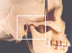
The temporomandibular joint (TMJ) is the joint between condylar head of the mandible and the mandibular fossa of the temporal bone. It is a synovial, condylar and hinge-type joint. The joint involves fibrocartilaginous surfaces and an articular disc which divides the joint into two cavities.[1] These superior and inferior articular cavities are lined by separate superior and inferior synovial membranes.[2] The TMJ is a part of the stomatognathic system. This system is made up off the TMJ, teeth and soft tissue. This system plays a role in breathing, eating and speech.[3] It also plays a role in kissing, yawning and sucking.[1]
Movements[edit | edit source]
A variety of movements occur at the TMJ. These movements are mandibular depression, elevation, lateral deviation (which occurs to both the right and left sides), retrusion and protrusion.
Muscles that act on the TMJ
| Muscles | Actions |
|---|---|
| Temporalis | Elevates mandible |
| Masseter | Elevates mandible |
| Lateral pterygoid | Protracts mandible, depresses chin, lateral deviation of mandible |
| Medial pterygoid | Works with masseter to elevate mandible, aids in protrusion, |
| Digastric
Stylohyoid Mylohyoid Geniohyoid |
Depresses the mandible against resistance when infrahyoid muscles stabilize or depress hyoid bone |
| Platysma | Depresses mandible against resistance |
Adapted from Moore[2]
Video showing movements of the TMJ[4]
Nerve Supply[edit | edit source]
The muscles that act on the TMJ are innervated by the mandibular nerve (CN V), the facial nerve (CN VII), C 1, C 2 and C 3.[2]
Resting Position and Close-Packed Position[edit | edit source]
The resting position of the TMJ is with the mouth slightly open, the lips together and the teeth not in contact. This is in contrast to the closed-pack position in which the teeth are tightly clenched.[1]
References[edit | edit source]
- ↑ 1.0 1.1 1.2 Magee DJ. Orthopedic physical assessment. 6th ed. Elsevier; 2014.
- ↑ 2.0 2.1 2.2 Moore KL, Dalley AF, Agur AM. Clinically oriented anatomy. Lippincott Williams & Wilkins; 2017 Sept 13.
- ↑ Di Fabio RP. Physical therapy for patients with TMD: a descriptive study of treatment, disability, and health status. Journal of orofacial pain. 1998 Apr 1;12(2).
- ↑ Functional Anatomy of the TMJ. Movements of the TMJ. Available from https://www.youtube.com/watch?v=SCS4MiHJ5Xw [last accessed 07/01/2018]


