Sternum
Original Editor - Grace Barla
Top Contributors - Grace Barla, Kim Jackson, Vaibhav Panchal, Lucinda hampton and Vidya Acharya
Description[edit | edit source]
The sternum is a flat cancellous bone with a compact cortex, it is slightly convex anteriorly, with multiple indentations along its lateral borders (costal notches). It forms the anterior median part of the thoracic skeleton. It is commonly known as the breastbone. In shape it resembles like a short sword. The sternum is located along the midline of body in the anterior thoracic region just deep to the skin. It is a flat bone about six inches in length, around an inch wide, and only a fraction of an inch thick. it is longer in males than in females.
Structure[edit | edit source]
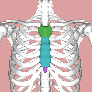
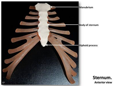
The sternum develops as three distinct parts:
- The manubrium
- The body of the sternum
- The xiphoid process
Manubrium
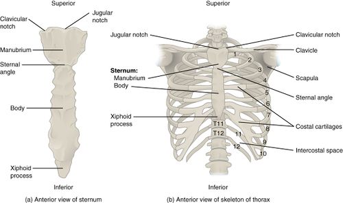
- Manubrium is at the cephalad end of the sternum is the large, flat, wide. At the superior border of the bone there is jugular notch or suprasternal notch. The clavicular notches for the articulation of clavicles are projected upward and laterally on both sides of jugular notch.
The manubrium makes angle with the body, convex forwards, called as the sternal angle of Louis. certain events take place at this angle -
- Formation of cardiac plexus
- Upper limit of base of heart
- Arch of aorta starts here as continuation of ascending aorta.
- Arch of aorta ends here to continue as descending thoracic aorta.
- Trachea divides into 2 branches.
The body
- It is the longest region of the sternum and is roughly rectangular in shape. It is also known as gladiolus. It has a convex anterior surface, and a concave posterior surface.
- It has facets on each side for articulation with the costal cartilage from third to seventh ribs with part of second costal cartilage.
Xiphoid process
- The sternal body joins the xiphoid process at the xiphosternal junction.
- It is a small projection of bone which is usually pointed.
- It varies greatly in shape and may be bifid or perforated.
- It lies in the floor of the epigastric fossa.
Function[edit | edit source]
Articulations[edit | edit source]
The cartilages from ribs 1 and 2 articulate with the manubrium, but the cartilage from rib 2 also articulates with the body of the sternum. The main articulations with the body of the sternum are with cartilages 2, 3, 4, 5, 6, and 7. The upward and outward movement of these cartilages causes the greatest increase in the internal diameter of the chest. The lateral projections of the sternum show where the clavicle and all the cartilages from the ribs articulate with the sternum itself. The sternal movement is greatest at this joint, the angulation between the manubrium and the body of the sternum is called as sternal angle. This angulation, forms the ‘‘bucket handle’’ that moves upward and outward, with the superior end of the manubrium being relatively fixed.
Muscle attachments[edit | edit source]
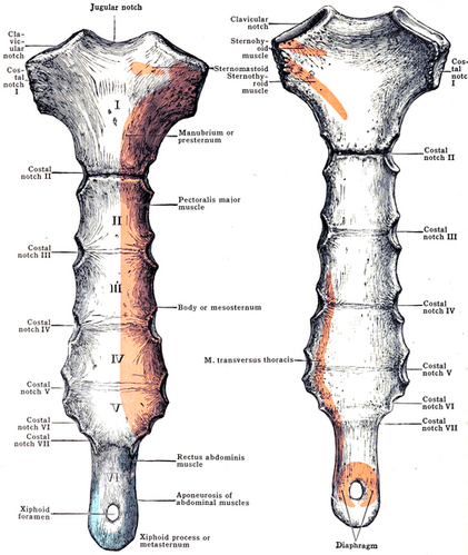
Manubrium The anterior surface gives origin on either side to:
- a. The pectoralis major.
- b. The sternal head of the sternocleidomastoid
The posterior surface gives origin to:
- a. The sternohyoid in upper part (Fig. 13.12).
- b. The sternothyroid in lower part.
The suprasternal notch gives attachment to the lower fibres of the interclavicular ligament, and to the two subdivisions of the investing layer of cervical fascia. The margins of each clavicular notch give attachment to the capsule of the corresponding sternoclavicular joint.
The body The anterior surface gives origin on either side to:
- the pectoralis major muscle.
The lower part of the posterior surface gives origin on either side to the sternocostalis muscle. Between the facets for articulation with the costal cartilages, the lateral borders provide attachment to:
- the external intercostal membranes
- internal intercostal muscles
The xiphoid The anterior surface provides insertion to the medial fibres of the rectus abdominis, and to the aponeuroses of the external and internal oblique muscles of the abdomen. The posterior surface gives origin to the diaphragm. The lateral borders of the xiphoid process give attachment to the aponeuroses of the internal oblique and transversus abdominis muscles. The upper end forms a primary cartilaginous joint with the body of the sternum. The lower end affords attachment to the linea alba.


