Search results
Page title matches

File:Activity One How do I use Es.png (785 × 132 (30 KB)) - 17:44, 27 January 2016
File:Activity Two How do I use ES.png (760 × 125 (22 KB)) - 17:52, 27 January 2016- 69 bytes (7 words) - 16:10, 4 July 2023
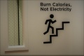
File:Image for sign to use stair 2.jpg (1,024 × 682 (121 KB)) - 23:33, 5 May 2018
File:Activity 3 How do I use ES.png (706 × 277 (54 KB)) - 18:27, 27 January 2016
File:Key Points How do I use ES.png (769 × 185 (47 KB)) - 18:32, 27 January 2016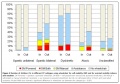
File:Wheelchair Use by Type of Cerebral Palsy.jpeg Rodby-Bousquet E, Hägglund G. Use of Manual and Powered Wheelchair in Children with Cerebral Palsy: A Cross-s {{fair-use}}(878 × 617 (154 KB)) - 00:41, 5 July 2018- ...nt. 2021 Jan 1;32:100296.</ref> Communities living in harsh conditions can use their mobile phone apps or platforms to: ...0163443718813486 The smartphone as a lifeline: An exploration of refugees’ use of mobile communication technologies during their flight.] Media, Culture &14 KB (1,917 words) - 12:24, 22 November 2023
File:Activity End of How do you use Es.png (761 × 242 (61 KB)) - 14:30, 28 January 2016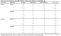
File:Use of Wheelchairs in Cerebral Palsy Related to Age.jpeg Rodby-Bousquet E, Hägglund G. Use of Manual and Powered Wheelchair in Children with Cerebral Palsy: A Cross-s {{fair-use}}(879 × 524 (96 KB)) - 00:42, 5 July 2018- 39 bytes (4 words) - 05:32, 6 December 2022
- 39 bytes (4 words) - 05:30, 6 December 2022
- == The use of a home program has many benefits for both the family and the child == ...n and intensity of using the home program is lacking.<ref name=":2" /> The use of these program in resource limited settings should also be investigated.12 KB (1,937 words) - 20:21, 5 April 2023
- ...ed these kinds of devices before, it can be a huge barrier to learn how to use them, before being able to experience the benefits they provide. ...in supporting these people, often they are eldery people, to learn how to use these devices and to benefit from them in their daily life.13 KB (2,005 words) - 12:08, 27 July 2022
- 69 bytes (7 words) - 22:56, 12 May 2020
- 69 bytes (7 words) - 16:12, 4 July 2023
- Acute Inpatient Rehabilitation Care for a Patient Learning to Use an above knee amputation (AKA) Prosthesis: Amputee Case Study ...lity in her home environment and to have the knowledge she needs to safely use her prosthesis. The patient was very clear she would only stay for 4-5 days11 KB (1,675 words) - 18:39, 21 February 2023
- '''Potential concerns related to the use of vibration therapy''' ...4(7):451-460. English, Spanish. </ref> note that research studies commonly use vague "expressions of perceived sensation", such as "below the motor thresh16 KB (2,171 words) - 00:33, 14 February 2023
- #REDIRECT [[Use of Hydrotherapy for the Management of Ankylosing Spondylitis (AS)]]83 bytes (11 words) - 20:03, 27 February 2023
- [[Hydrotherapy]] is the external or internal use of water in any of its form (water, ice, steam) for health promotion or tre ==== Simple Use of Running Water ====36 KB (5,296 words) - 20:03, 27 February 2023


Page text matches
File:Deep flexor muscles of the forearm Primal.png ...ven permission to use this image exclusively in Physiopedia. Please do not use this image outside Physiopedia unless you have prior permission(660 × 660 (189 KB)) - 00:25, 7 December 2020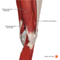
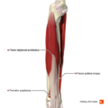
File:Intermediate muscles of the knee posterolateral aspect Primal.png ...ven permission to use this image exclusively in Physiopedia. Please do not use this image outside Physiopedia unless you have prior permission(660 × 660 (342 KB)) - 21:39, 7 December 2020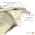
File:Acromioclavicular separation type 1 Primal.png ...ven permission to use this image exclusively in Physiopedia. Please do not use this image outside Physiopedia unless you have prior permission(660 × 660 (274 KB)) - 01:08, 8 December 2020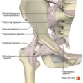
File:Ligaments of the hip joint anterior aspect Primal.png ...ven permission to use this image exclusively in Physiopedia. Please do not use this image outside Physiopedia unless you have prior permission(660 × 660 (361 KB)) - 20:29, 7 December 2020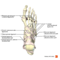
File:Ligaments of the foot dorsal aspect Primal.png ...ven permission to use this image exclusively in Physiopedia. Please do not use this image outside Physiopedia unless you have prior permission(660 × 660 (230 KB)) - 00:20, 8 December 2020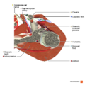
File:Axial section of the shoulder joint Primal.png ...ven permission to use this image exclusively in Physiopedia. Please do not use this image outside Physiopedia unless you have prior permission(990 × 990 (707 KB)) - 01:15, 8 December 2020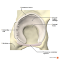
File:Radial fibrillated labral tear Primal.png ...ven permission to use this image exclusively in Physiopedia. Please do not use this image outside Physiopedia unless you have prior permission(990 × 990 (539 KB)) - 20:56, 7 December 2020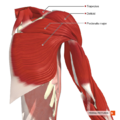
File:Muscles connecting the upper limb to the trunk anterior aspect Primal.png ...ven permission to use this image exclusively in Physiopedia. Please do not use this image outside Physiopedia unless you have prior permission(660 × 660 (485 KB)) - 00:58, 8 December 2020
File:Posterior disc hernia sagittal view Primal.png ...ven permission to use this image exclusively in Physiopedia. Please do not use this image outside Physiopedia unless you have prior permission(990 × 990 (660 KB)) - 15:21, 8 December 2020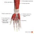
File:Muscles of the hand anterior aspect Primal.png ...ven permission to use this image exclusively in Physiopedia. Please do not use this image outside Physiopedia unless you have prior permission(660 × 660 (265 KB)) - 00:26, 7 December 2020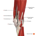
File:Intermediate muscles of the knee posteromedial aspect Primal.png ...ven permission to use this image exclusively in Physiopedia. Please do not use this image outside Physiopedia unless you have prior permission(660 × 660 (355 KB)) - 21:40, 7 December 2020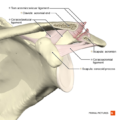
File:Acromioclavicular separation type 2 Primal.png ...ven permission to use this image exclusively in Physiopedia. Please do not use this image outside Physiopedia unless you have prior permission(660 × 660 (287 KB)) - 01:09, 8 December 2020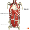
File:Anterior abdominal wall deep muscles Primal.png ...ven permission to use this image exclusively in Physiopedia. Please do not use this image outside Physiopedia unless you have prior permission(660 × 660 (412 KB)) - 21:47, 8 December 2020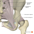
File:Ligaments of the hip joint posterior aspect Primal.png ...ven permission to use this image exclusively in Physiopedia. Please do not use this image outside Physiopedia unless you have prior permission(660 × 660 (395 KB)) - 20:40, 7 December 2020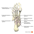
File:Ligaments of the foot plantar aspect Primal.png ...ven permission to use this image exclusively in Physiopedia. Please do not use this image outside Physiopedia unless you have prior permission(660 × 660 (234 KB)) - 00:22, 8 December 2020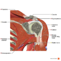
File:Coronal section of the tendon of long head of biceps Primal.png ...ven permission to use this image exclusively in Physiopedia. Please do not use this image outside Physiopedia unless you have prior permission(990 × 990 (900 KB)) - 01:17, 8 December 2020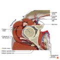
File:Axial section of the hip joint Primal.png ...ven permission to use this image exclusively in Physiopedia. Please do not use this image outside Physiopedia unless you have prior permission(990 × 990 (881 KB)) - 20:57, 7 December 2020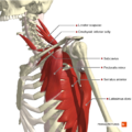
File:Muscles connecting the upper limb to the trunk deep muscles Primal.png ...ven permission to use this image exclusively in Physiopedia. Please do not use this image outside Physiopedia unless you have prior permission(660 × 660 (464 KB)) - 00:59, 8 December 2020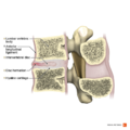
File:Anterior disc hernia sagittal view Primal.png ...ven permission to use this image exclusively in Physiopedia. Please do not use this image outside Physiopedia unless you have prior permission(990 × 990 (660 KB)) - 15:23, 8 December 2020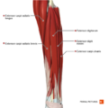
File:Superficial and intermediate extensor muscles of the forearm Primal.png ...ven permission to use this image exclusively in Physiopedia. Please do not use this image outside Physiopedia unless you have prior permission(660 × 660 (236 KB)) - 00:27, 7 December 2020


