Sinus Tarsi Syndrome
Original Editors - Merlin Roggeman
Lead Editors - Your name will be added here if you are a lead editor on this page. Read more.
Search Strategy[edit | edit source]
To search for information you can use databases such as PubMed, Web of Science, PEDro or Google Scholar. Use keywords as ‘sinus tarsi syndrome’ or ‘sinus tarsitis’. For specific information you can use ‘AND’ and add ‘physical therapy’, ‘characteristics’, ‘definition’, depending on what you search. Also choose articles from which the full text is available for free.
Definition/Description[edit | edit source]
The sinus tarsi syndrome is a foot pathology, mostly following after a traumatic injury to the ankle. It may also occur if the person has a pes planus or an (over)-pronated foot, which can cause compression in the sinus tarsi. Some characteristics are pain at the lateral side of the ankle and a feeling of instability. [1][2]
Clinically Relevant Anatomy[edit | edit source]
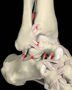
The sinus tarsi is a tunnel between the talus and the calcaneus, which contains some anatomic structures that can be injured in STS. Some ligaments founding this region are the interosseus talo-calcaneal ligament (number 5 in figure 1), the cervical ligament (number 6 in figure 1), and the medial, lateral and intermediate roots of the inferior extensor retinaculum.[1][2][3][4]
The sural nerve passes the sinus tarsi laterally, and some branches of it may run through the tarsi.[4]
Figure 1: an image of the ankle, with the sinus tarsi between the talus and calcaneus, and the ligaments in the sinus (numbers 5 and 6) (source: http://www.blackburnfeet.org.uk/hyperbook/trauma/ankleFx/ankleFxBasic1.htm)
Epidemiology /Etiology[edit | edit source]
Mostly the sinus tarsi syndrome occurs after a traumatic lateral ankle sprain. The ligaments of the sinus tarsi can be sprained or torn, and an inflammation and hemorrhage of the synovial recess in the sinus tarsi can occur. This happens in 70% of the cases.[1][2][4]
The sinus tarsi syndrome can also occur as a compression injury, for example to people who have flat or pronated feet. The talus and calcaneus are pressed together as a result of the deformation. This causes bone to bone contact of the talus and calcaneus, with inflammation or arthritis in the sinus.[1][4]
Characteristics/Clinical Presentation[edit | edit source]
The characteristics of the syndrome are pain at the lateral side of the ankle. “The pain is most severe when standing, walking on uneven ground or during the movements of supination and adduction of the foot”[1] People suffering from the sinus tarsi syndrome also have a feeling of instability (functional instability) in the hind foot.[1][4]
When the syndrome is a result of an inverted ankle sprain there is a major chance the lateral collateral ligaments of the ankle are also damaged, since the ligaments in the sinus tarsi are the last ones to tear with a traumatic ankle sprain.[1][4]
Differential Diagnosis[edit | edit source]
These common pathologies may give the same pain characteristics or symptoms:[5]
-ankle sprain
-calcaneal fracture
-talar fracture
-peroneal tendonitis
-subtalar joint arthritis
-tarsal tunnel syndrome
Diagnostic Procedures[edit | edit source]
Diagnosis of the sinus tarsi syndrome is usually made by excluding other foot pathologies. CT-scans exclude bone fractures, but are not specific enough to diagnose STS. The most commonly used methods are MRI’s. MRI findings may include filling of the sinus tarsi space with fluid or scar tissue, alterations in the structure of the ligaments or degenerative changes in the subtalar joint.[2][3][6]
Outcome Measures[edit | edit source]
add links to outcome measures here (also see Outcome Measures Database)
Examination[edit | edit source]
When having a patient with pain in the lateral ankle, the physiotherapist should check eventual functional instability and compensation with the other ankle.[2]
In standing posture the patients may demonstrate a pes planus posture or an asymmetry of the rearfoot angle with the leg.[2]
In passive examination, the range of motion of the ankle may be limited in pronation and supination, but pain over the sinus tarsi at the end range of plantar flexion combined with supination is a typical sign for STS. The subtalar joint may have increased translation mobility if the interosseus and cervical ligaments are disrupted, but this is not always the case.[2]
The therapist should also evaluate if there is any muscle weakness of the peroneal and planatr flexor muscles. This is done with resistance tests of the ankle: pronation tests and flexion tests.[2][4]
Medical Management
[edit | edit source]
The treatment of the sinus tarsi syndrome can be conservative or operative.
The first one includes physiotherapy (see physical therapy management), injections with corticosteroids in the sinus tarsi, local gels or drugs.[1][4]
Operative treatment is also very effective in most cases, but needs to be considered as a last resort if conservative treatment fails.[4]
Physical Therapy Management
[edit | edit source]
add text here
Key Research[edit | edit source]
add links and reviews of high quality evidence here (case studies should be added on new pages using the case study template)
Resources
[edit | edit source]
add appropriate resources here
Clinical Bottom Line[edit | edit source]
add text here
Recent Related Research (from Pubmed)[edit | edit source]
see tutorial on Adding PubMed Feed
Extension:RSS -- Error: Not a valid URL: Feed goes here!!|charset=UTF-8|short|max=10
References[edit | edit source]
see adding references tutorial.
- ↑ 1.0 1.1 1.2 1.3 1.4 1.5 1.6 1.7 Taillard W, Meyer JM, Garcia J, Blanc Y. The Sinus Tarsi Syndrome. International Orthopaedics (SICOT) 1981; 5:117-130
- ↑ 2.0 2.1 2.2 2.3 2.4 2.5 2.6 2.7 Helgeson K. Examination and Intervention for Sinus Tarsi Syndrome. North American Journal of Sports Physical Therapy 2009 February; 4(1):29-37
- ↑ 3.0 3.1 Rosenberg ZS, Beltran J, Bencardino JT. From the RSNA Refrecher Courses. MR Imaging of the Ankle and Foot. RadioGraphics 2000; 20:153-179
- ↑ 4.0 4.1 4.2 4.3 4.4 4.5 4.6 4.7 4.8 Meir Nyska, Gideon Mann, editors. The unstable ankle. Chapter 14: Sinus Tarsi Syndrome. United States: Human Kinetics Publishers, Inc. 2002. p144-120
- ↑ MyFootShop. Sinus Tarsi Syndrome. http://www.myfootshop.com/detail.asp?Condition=Sinus%20Tarsi%20Syndrome (accessed 28/11/2010)
- ↑ American Academy of Podiatric Sports Medicine. Sinus Tarsi Syndrome. http://www.aapsm.org/sinus_tarsi_syndrome.html (accessed 27/11/2010)


