Scaphoid: Difference between revisions
Nina Myburg (talk | contribs) (Content) |
Nina Myburg (talk | contribs) (Added images) |
||
| Line 5: | Line 5: | ||
</div> | </div> | ||
== Description == | == Description == | ||
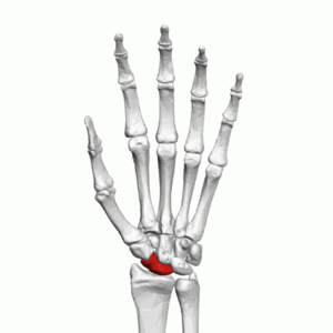
The scaphoid is the largest bone of the proximal row of carpal bones.<ref name=":1">Moore KL, Dalley AF. ''Clinically Oriented Anatomy.'' Fifth edition. Philadelphia: Lippincot Williams & Wilkins; 2006</ref> The word scaphoid is derived from the Greek word ''skaphos'' which means "a boat". The name refers to the boat-like shape of the bone. Previously it was called the navicular bone (derived from the Latin word ''navis'' which means "boat") of the hand. However, there is a tarsal bone in the foot that is also called navicular due to its boat-like shape. The scaphoid is situated at the radial side of the wrist.<ref name=":0">Gray H. ''[https://www.bartleby.com/107/54.html Anatomy of the Human Body].'' Twentieth edition. Philadelphia: Lea & Febiger; 1918 Available from: https://www.bartleby.com/107/54.html [Accessed 19 June 2019]</ref> | The scaphoid is the largest bone of the proximal row of carpal bones.<ref name=":1">Moore KL, Dalley AF. ''Clinically Oriented Anatomy.'' Fifth edition. Philadelphia: Lippincot Williams & Wilkins; 2006</ref> The word scaphoid is derived from the Greek word ''skaphos'' which means "a boat". The name refers to the boat-like shape of the bone. Previously it was called the navicular bone (derived from the Latin word ''navis'' which means "boat") of the hand. However, there is a tarsal bone in the foot that is also called navicular due to its boat-like shape. The scaphoid is situated at the radial side of the wrist.<ref name=":0">Gray H. ''[https://www.bartleby.com/107/54.html Anatomy of the Human Body].'' Twentieth edition. Philadelphia: Lea & Febiger; 1918 Available from: https://www.bartleby.com/107/54.html [Accessed 19 June 2019]</ref> | ||
[[File:Scaphoid bone (left hand) - animation.gif|center|thumb|Scaphoid bone (animation) - Left hand]] | |||
== Structure == | == Structure == | ||
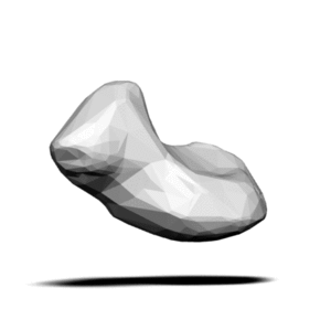
The scaphoid is situated below the radius at the radial (lateral) side of the wrist. It has a boat-like shape with many articulation surfaces. Over 80% of the bone is covered in cartilage. On the palmar surface of the bone, there is a tubercle that can be palpated at the base and medial aspect of the thenar eminence when the hand is extended. <ref name=":1" /> It can also be palpated at the base of the anatomical snuffbox. | The scaphoid is situated below the radius at the radial (lateral) side of the wrist. It has a boat-like shape with many articulation surfaces. Over 80% of the bone is covered in cartilage. On the palmar surface of the bone, there is a tubercle that can be palpated at the base and medial aspect of the thenar eminence when the hand is extended. <ref name=":1" /> It can also be palpated at the base of the anatomical snuffbox. | ||
[[File:Scaphoid bone (picture of only the bone) - animation1.gif|center|thumb|Scaphoid bone]] | |||
== Function == | == Function == | ||
Revision as of 12:05, 19 June 2019
Original Editor
Top Contributors - Nina Myburg, Kim Jackson, Chrysolite Jyothi Kommu, Nikhil Benhur Abburi and Vidya Acharya
Description[edit | edit source]
The scaphoid is the largest bone of the proximal row of carpal bones.[1] The word scaphoid is derived from the Greek word skaphos which means "a boat". The name refers to the boat-like shape of the bone. Previously it was called the navicular bone (derived from the Latin word navis which means "boat") of the hand. However, there is a tarsal bone in the foot that is also called navicular due to its boat-like shape. The scaphoid is situated at the radial side of the wrist.[2]
Structure[edit | edit source]
The scaphoid is situated below the radius at the radial (lateral) side of the wrist. It has a boat-like shape with many articulation surfaces. Over 80% of the bone is covered in cartilage. On the palmar surface of the bone, there is a tubercle that can be palpated at the base and medial aspect of the thenar eminence when the hand is extended. [1] It can also be palpated at the base of the anatomical snuffbox.
Function[edit | edit source]
The scaphoid, together with other carpal bones, provide bony structure to the hand and wrist. It is involved in movement of the wrist together with the lunate and distal surfaces of the radius and ulna. It forms the radial border of the carpal tunnel and serves as attachment site.
Articulations[edit | edit source]
The superior surface is convex, smooth, of triangular shape, and articulateswith the lower end of the radius. The inferior surface, directed downward, lateralward, and backward, is also smooth, convex, and triangular, and is divided by a slight ridge into two parts, the lateral articulating with the greater multangular, the medial with the lesser multangular. On the dorsal surface is a narrow, rough groove, which runs the entire length of the bone, and serves for the attachment of ligaments. The volar surface is concave above, and elevated at its lower and lateral part into a rounded projection, the tubercle, which is directed forward and gives attachment to the transverse carpal ligament and sometimes origin to a few fibers of the Abductor pollicis brevis. The lateral surface is rough and narrow, and gives attachment to the radial collateral ligament of the wrist. The medial surface presents two articular facets; of these, the superior or smaller is flattened of semilunar form, and articulates with the lunate bone; the inferior or larger is concave, forming with the lunate a concavity for the head of the capitate bone[2]
The palmar surface of the scaphoid is concave, and forming a tubercle, giving attachment to the transverse carpal ligament. The proximal surface is triangular, smooth and convex, and articulates with the radius and adjacent carpal bones, namely the lunate, capitate, trapezium and trapezoid.[2] The lateral surface is narrow and gives attachment to the radial collateral ligament. The medial surface has two facets, a flattened semi-lunar facet articulating with the lunate bone, and an inferior concave facet, articulating alongside the lunate with the head of the capitate bone.[3]
The dorsal surface of the bone is narrow, with a groove running the length of the bone and allowing ligaments to attach, and the surface facing the fingers (anatomically inferior) is smooth and convex, also triangular, and divided into two parts by a slight ridge.[3]
The navicular articulates with five bones: the radius proximally, greater and lesser multangulars distally, and capitate and lunate medially.[2]
It is located on the radial side of the wrist, and articulates with the radius, lunate, trapezoid, trapezium and capitate
The superior surface is convex, smooth, of triangular shape, and articulateswith the lower end of the radius. The inferior surface, directed downward, lateralward, and backward, is also smooth, convex, and triangular, and is divided by a slight ridge into two parts, the lateral articulating with the greater multangular, the medial with the lesser multangular. On the dorsal surface is a narrow, rough groove, which runs the entire length of the bone, and serves for the attachment of ligaments. The volar surface is concave above, and elevated at its lower and lateral part into a rounded projection, the tubercle, which is directed forward and gives attachment to the transverse carpal ligament and sometimes origin to a few fibers of the Abductor pollicis brevis. The lateral surface is rough and narrow, and gives attachment to the radial collateral ligament of the wrist. The medial surface presents two articular facets; of these, the superior or smaller is flattened of semilunar form, and articulates with the lunate bone; the inferior or larger is concave, forming with the lunate a concavity for the head of the capitate bone[2]
Soft tissue attachments[edit | edit source]
Clinical relevance[edit | edit source]
Clicking of the scaphoid or no anterior translation can indicate scapholunate instability
See also[edit | edit source]
References[edit | edit source]
- ↑ 1.0 1.1 Moore KL, Dalley AF. Clinically Oriented Anatomy. Fifth edition. Philadelphia: Lippincot Williams & Wilkins; 2006
- ↑ 2.0 2.1 2.2 2.3 Gray H. Anatomy of the Human Body. Twentieth edition. Philadelphia: Lea & Febiger; 1918 Available from: https://www.bartleby.com/107/54.html [Accessed 19 June 2019]


