Rubrospinal Tract
Original Editor - Kate Sampson
Lead Editors - Kate Sampson, Lucinda hampton, Evan Thomas, Wendy Walker, WikiSysop and Kim Jackson
Description[edit | edit source]
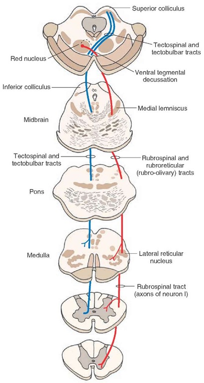
The Rubrospinal tract is a descending pathway which originates in the Red Nucleus and descends to the spinal cord.[1]
Anatomy[edit | edit source]
 Origin[edit | edit source]

- The Red Nucleus of the midbrain tegmentum[1]
Course / Path[edit | edit source]
- Fibres pass ventromedially and cross the ventral tegmental decussation [1]
- Fibres descend to the spinal cord where they lie ventrolateral to and intertwined with the corticospinal tract [1]
Function[edit | edit source]
- As the Rubrospinal tract receives afferent fibres from the motor cortex and cerebellum it acts as a non pyramidal route of influencing spinal cord activity through inter and motor neurones [1][2]
- Excitation of the motor neurons controlling tone of limb flexor muscles and inhibitory to extension during of gait[1][3]
- Facilitatory of flexion and inhibitory to extension in cervical and lumbar spine and distal extremity muscles[4]
- Neural activity in the red nucleus is related to force, velocity and direction of movement[5]
Pathology[edit | edit source]
Within the literature, selective lesions have not been reported of solely the red nucleus or rubrospinal tract. Lesions within the region of the red nucleus can result in movement disorders and tremor, but these effects may be more associated with damage to fibers which are associated with the cerebellar and basal ganglia systems.[6]
In primate studies, it has been suggested that the rubrospinal tract is responsible for fractionation of movement.[7]. Fractionation is the ability to isolate movement to one joint independent of another.[8] Therefore, it could be hypothesised that if the rubrospinal tract was affected, this could have an impact on the fine tuning and fractionation of movement.
However, even though it has been shown within animal studies that the rubrospinal tract can assist with functional recovery, it is unclear as to how this can be generalised to humans.[9]
References[edit | edit source]
- ↑ 1.0 1.1 1.2 1.3 1.4 1.5 Crossman AR, Neary D. Neuroanatomy: An Illustrated Colour Text. Third Edition. London: Elsevier, 2004
- ↑ Martinez-Lopez1 JE, Moreno-Bravo1 JA, Madrigal1 MP, Martinez1 S, Puelles1 E. Red nucleus and rubrospinal tract disorganization in the absence of Pou4f1. Front. Neuroanat 2015;9:8.
- ↑ Kidd G, Lawes N, Musa I. Understanding Neuromuscular Plasticity. London: Edward Arnold, 1992.
- ↑ Rothwell J. Control of Human Voluntary Movement. London: Chapman & Hall, 1994
- ↑ Leonard CT. The Neuroscience of Human movement. St Louis: Mosby 1998
- ↑ What-When-How. The Upper Motor Neurons (Motor Systems) Part 3. http://what-when-how.com/neuroscience/the-upper-motor-neurons-motor-systems-part-3/ (accessed on 1/4/2016)
- ↑ Belhaj-Saif A, Cheney PD. Plasticity in the distribution of the red nucleus output to forearm muscles after unilateral lesions of the pyramidal tract. J Neurophysiol 2000; 83: 3147–53
- ↑ Scheets PL, Sahrmann SA, Norton BJ. Use of movement system diagnoses in the management of patients with neuromuscular conditions: a multiple-patient case report. Physical therapy. 2007 May 15.
- ↑ Onodera S, Hicks TP. Carbocyanine dye usage in demarcating boundaries of the aged human red nucleus. PloS one 2010; 5: e14430.

