Reticulospinal Tract: Difference between revisions
Kate Sampson (talk | contribs) No edit summary |
Kate Sampson (talk | contribs) No edit summary |
||
| Line 46: | Line 46: | ||
*Responsible for flexor motor neurons | *Responsible for flexor motor neurons | ||
*Inhibits the medial reticulospinal tract and therefore extensor motor neurones <ref name="Trompetto" /> | *Inhibits the medial reticulospinal tract and therefore extensor motor neurones enabling modulation of the stretch reflex <ref name="Trompetto" /> | ||
==== Locomotion ==== | ==== Locomotion ==== | ||
Revision as of 20:04, 6 May 2016
Original Editor - Kate Sampson
Top Contributors - Kate Sampson, Lucinda hampton, 127.0.0.1, Wendy Walker, Evan Thomas, WikiSysop and Kim Jackson
Description[edit | edit source]
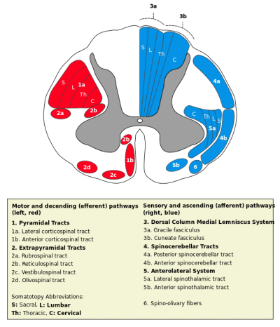
The Reticulospinal tract is responsible primarily for locomotion and postural control. The Reticulospinal tract is comprised of the medial (pontine) tract and the lateral (medullary) tract.[1]
Anatomy[edit | edit source]
Origin[edit | edit source]
- Reticular formation in the pontine (Medial Reticulospinal tract) and medulla (Lateral Reticulospinal tract)[2]
Course / Path[edit | edit source]
Medial Reticulospinal Tract (Pontine)[edit | edit source]
- Descends ipsilaterally in the anterior funiculus
Lateral Reticulospinal tracts (Medullary)[edit | edit source]
- Descends bilaterally in the lateral funiculus
Both the lateral and medial tracts act via internuncials shared with the corticospinal tract on proximal limb and axial muscle motor neurons.[1]
Function[edit | edit source]
- Control the activity of both alpha and gamma motor neurones[2]
- Mediate pressor and depressor effects on the circulatory system[2]
- Help to control breathing[2]
- Due to the interrelation between vestibulospinal and ipsilateral reticulospinal tracts, this can result in selective activation of many muscles at the same time
- As they work through interneurons and long propriospinal neurons, they can enable co-ordinated, selective movements[3]
- The relationship between the rubrospinal and crossed reticulospinal tracts can result in a postural role within distal musculature
- The pathways innervate motorneurons both directly and indirectly through interneurons and short propriospinal neurons (e.g. intrinsic muscles acting in a postural role for individual finger movement)
Medial Reticulospinal Tract (Pontine)[edit | edit source]
- Responsible for controlling extensor motor neurons
- Stimulation of the midbrain locomotor centre can result patterned movements (e.g. stepping)[3]
Lateral Reticulospinal Tract (Medullary)[edit | edit source]
- Responsible for flexor motor neurons
- Inhibits the medial reticulospinal tract and therefore extensor motor neurones enabling modulation of the stretch reflex [4]
Locomotion[edit | edit source]
When generating movements in two sides of the body within humans, it results in reciprocal inhibition of the flexors and extensors.
In animal studies, central pattern generators (CPGs) are found to be responsible for locomotion generation. As locomotion is generated by internuncial neurons in the cervical and lumbar regions, activating the flexors and extensors, the intermediate gray matter is able to initiate rhythmical movements.[1]
In humans, locomotion is generated in the locomotion centre in the midbrain. The premotor cortex which is responsible for the overarching control of locomotion projects to the brainstem and therefore reticulospinal tract. As a consequence, the reticulospinal tract is able to modulate control of locomotion.[1] The reticular formation, due to its role in attention and conscious perception, can result in a specific reaction to sensory information. The cortico-reticulospinal tract is thereby responsible for transmitting excitatory and inhibitory information to be processed before being passed to the spinal cord.[3]
Posture[edit | edit source]
The reticular formation within the pons is partly responsible for postural control functions.
The premotor cortex is able to identify appropriate axial musculature to enable distal movement.[1] Due to the interrelation of internuncial neurons between the corticospinal and reticulospinal tracts, either the pyramidal or extrapyramidal tracts can be responsible for controlling movement patterns. The extrapyramidal system (reticulospinal) tract can initiate movement within a stable, routine situation, whereas the pyramidal (corticospinal) tract is able to control tasks requiring more cognitive appraisal (e.g. varying surface, distractions and requiring close attention).
Pathology[edit | edit source]
Lesions to the cortico-reticulospinal system can result in decreased postural control and reduced selectivity of postural control.[3]
Also due to the lateral reticulospinal's role in inhibition, if this pathway is disruped it can result in spasticity [4]. In addition due to the lack of descending inhibition, the medial reticulospinal tract then maintains spasticity in the musculature.[4]
Recent Related Research (from Pubmed)[edit | edit source]
Failed to load RSS feed from http://www.ncbi.nlm.nih.gov/entrez/eutils/erss.cgi?rss_guid=1hIuyCTdZ6akVq09ES1z0X4jYomaY_M3XcXsbDFXyYku2mns9F|charset=UTF-8|short|max=10: Error parsing XML for RSS
References[edit | edit source]
- ↑ 1.0 1.1 1.2 1.3 1.4 Fitzgerald MJT, Gruener G, Mtui E. Clinical neuroanatomy and neuroscience. Fifth Edition. Philadelphia: Elsevier Saunders, 2007
- ↑ 2.0 2.1 2.2 2.3 Crossman AR, Neary D. Neuroanatomy. An Illustrated colour text. Third Edition. Philadelphia: Churchill Livingstone, 2005
- ↑ 3.0 3.1 3.2 3.3 Gjelsvik BEB. The bobath concept in adult neurology. Stuttgart: Thieme, 2008
- ↑ 4.0 4.1 4.2 Trompetto C, Marinelli L, Mori L, Pelosin E, Currà A, Molfetta L, Abbruzzese G. Pathophysiology of spasticity: Implications for neurorehabilitation. BioMed research international. 2014 Oct 30;2014.


