Posterior atlanto-occipital ligament: Difference between revisions
No edit summary |
Kim Jackson (talk | contribs) m (Text replacement - "Category:Anatomy - Cervical Spine" to "") |
||
| (12 intermediate revisions by 4 users not shown) | |||
| Line 1: | Line 1: | ||
<div class="editorbox"> | <div class="editorbox"> | ||
'''Original Editor '''- | '''Original Editor '''- [[User:Rachael Lowe|Rachael Lowe]] | ||
'''Top Contributors''' - {{Special:Contributors/{{FULLPAGENAME}}}} | '''Top Contributors''' - {{Special:Contributors/{{FULLPAGENAME}}}} | ||
</div> | </div> | ||
== Description == | == Description == | ||
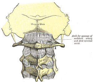
[[Image:Atlanto-occipital joint posterior.png|thumb|right|Posterior atlanto-occipital ligament and membrane]] | |||
The posterior atlantooccipital membrane (posterior atlantooccipital ligament) is a broad but thin membrane. It is connected above to the posterior margin of the foramen magnum and below to the upper border of the posterior arch of the atlas. It is a continuation from the Ligamentum Flavum. | |||
On each side of this membrane there is defect above the groove for the vertebral artery which serves as an opening for the entrance of the artery. The suboccipital nerve also passes through this defect. | |||
The membrane | The free border of the membrane arches over the artery and nerve and is sometimes ossified. | ||
The membrane is deep to the [[Rectus Capitis Posterior Minor|rectus capitis posterior minor]] and [[Obliquus Capitis Superior|obliquus capitis superior]] muscles, and is superficial to the dura mater of the vertebral canal to which it is closely associated. | |||
== References == | == References == | ||
<references /><br> | |||
[[Category:Anatomy]] | |||
[[Category:Cervical Spine]] | |||
[[Category:Ligaments]] | |||
[[Category:Musculoskeletal/Orthopaedics]] | |||
[[Category:Cervical Spine - Ligaments]] | |||
Latest revision as of 13:40, 23 August 2019
Original Editor - Rachael Lowe
Top Contributors - Admin, Kim Jackson, Evan Thomas and WikiSysop
Description[edit | edit source]
The posterior atlantooccipital membrane (posterior atlantooccipital ligament) is a broad but thin membrane. It is connected above to the posterior margin of the foramen magnum and below to the upper border of the posterior arch of the atlas. It is a continuation from the Ligamentum Flavum.
On each side of this membrane there is defect above the groove for the vertebral artery which serves as an opening for the entrance of the artery. The suboccipital nerve also passes through this defect.
The free border of the membrane arches over the artery and nerve and is sometimes ossified.
The membrane is deep to the rectus capitis posterior minor and obliquus capitis superior muscles, and is superficial to the dura mater of the vertebral canal to which it is closely associated.
References[edit | edit source]


