Popliteus strain
This article is currently under review and may not be up to date. Please come back soon to see the finished work! (16/04/2022)
Original Editors - Melissa Decoen as part of the Vrije Universiteit Brussel's Evidence-based Practice project
Top Contributors - Erika Rodrigues, Hardik Bhatt, Melissa Decoen, Kim Jackson, Admin, Claire Knott, Wanda van Niekerk, Daphne Jackson, WikiSysop, Evan Thomas and Oyemi Sillo
Introduction[edit | edit source]
Popliteus muscle injuries are rarely isolated injuries and are mostly associated with other injuries of the posterolateral corner of the knee such as lateral collateral ligament injury, Anterior cruciate ligament injury , posterior cruciate ligament injury or meniscal tear.[1]
Clinically Relevant Anatomy[edit | edit source]
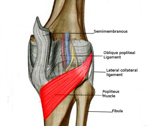
The Popliteus Muscle tendinous unit is unique in that the distal muscular attachment is designated the insertion and the tendinous proximal (femoral) attachment is designated the origin. The muscle inserts into a triangular area along the posteromedial aspect of the proximal tibial metaphysic above the soleal line. It forms the floor of the popliteus fossa. The tendon of the popliteus passes through the popliteal hiatus, entering the knee joint and inserting into the lateral femoral condyle at the end of the popliteal sulcus. The main tendinous component inserts into the lateral femoral condyle with variable aponeurotic attachments to the posterior horn of the lateral meniscus and the fibular head[2]. The insertion into the lateral meniscus retracts and protects the meniscus in flexion, but this function has been disputed. The femoral insertion has a crescent shape, with the superior aspect being concave.[3] The main tendon of the popliteus muscle consists of anterior and posterior fibers.[4] The popliteus muscle is innervated by tibial nerves (L4-L5 and S1).The popliteus tendon is intra-capsular but extra-articular and extra synovial.[5]
Functions of the Politeus Muscle at the Knee joint[edit | edit source]
The popliteus muscle has both static and dynamic function at the knee joint.
Dynamic functions : During initial flexion of the knee, it initiates and maintains the internal rotation of the tibia on the femur (unlocking of knee joint) and prevents forward dislocation of the femur on the tibia.
It acts as a secondary restraint to posterior displacement of the tibia in PCL deficient knees and is an important Static Stabilizer of the posterolateral corner of the knee. [2]
Mechanism of Injury[edit | edit source]
- Contact/ Traumatic Injuries - Forced external rotation of the tibia on a partially flexed knee, falls onto an extended knee or forced hyperextension.[1]
- Non-Contact/Non-Traumatic Injuries - Forced external rotation of the tibia with an associated varus force applied to a fixed tibia[1]
Clinical Presentation[edit | edit source]
- Pain over the lateral joint line
- Pain at the back of the knee
- Pain on weight-bearing and stair-climbing
- Incomplete Range of motion
Types of Injuries[edit | edit source]
- Popliteus Tendon Avulsion
- Popliteus Tendon Tear (Partial/ Complete)
Differential Diagnosis[edit | edit source]
- Biceps femoris tendon strain[6]
- Lateral meniscus Injury
- Lateral collateral ligament injury
- Tibial nerve injury
Diagnostic Procedures[edit | edit source]
Complete tears of muscle can be clinically identified however the popliteus muscle is an exception. It is difficult to identify which structure of posterolateral complex is injured and therefore MRI is the preferred diagnostic tool.
An acute haemarthrosis and lateral pain in a stable knee should lead to suspicion of an isolated injury to the popliteus muscle-tendon unit.[7] The diagnosis should be entertained in any acutely swollen knee with posterolateral tenderness and pain on resisted internal tibial rotation.[8]
MRI of the knee should be performed to evaluate the nature of the injury. The diagnosis may be confirmed by arthroscopic examination of the knee.
In some cases, the ruptured popliteus tendon retracts distally through the popliteal hiatus and can no longer be seen in the joint[9][10][11].
Diagnostic Test[edit | edit source]
1. Garrick Test
Patient seated, hip and knee are both flexed to 90°. The patient actively externally rotates the lower leg and this is resisted by the examiner. A positive test is pain during the maneuver in the location of the popliteus muscle or tendon.
2. Shoe Removal Maneuver
This test is where pain is felt when trying to remove the shoe on the other side of the affected knee. This motion requires internal rotation of the affected leg to reach the heel of the other leg and can cause pain during the maneuver when there is injury to the popliteus muscle.
Knee outcome survey can be used as an outcome measure
Examination[edit | edit source]
Clinically the affected patients present with an unnatural outward rotation of the tibia when bending the knee. Additionally, other general symptoms often occur such as muscle swelling, edema or bleeding[12]
Only a portion of the popliteus can be safely palpated due to the neurovascular structures that overlie it. The attachment on the tibial shaft can usually be reached as well as the tendon at the femoral condyle.
Rest described above in diagnostic tests.
| POSTERIOR KNEE JOINT ASSESMENT |
|---|
| Patient should be assessed in supine and prone lying. Pain or ‘a feeling of fullness’ in the posterior knee can be the sign of a joint effusion. |
| Active and passive range of movement of the knee joint should be assessed in flexion, extension and tibio-femoral rotation. |
| Manual muscle testing of knee flexion should be performed with the knee joint positioned in both neutral and external rotation. |
| Palpation of the joint line, the tendons of the hamstrings, gastrocnemius and the popliteus should be carried out. |
| The posterior draw tests to check the integrity of the Posterior cruciate ligament. |
| Posterolateral rotational stability of the knee should be checked using either the recurvatum test , reverse pivot shift or dial test. Isolated popliteus injuries do not cause significant posterolateral rotational instability as compared to injuries to the posterolateral corner of the knee. |
Medical Management[edit | edit source]
Medical management as line with other muscle injuries as it is mostly associate with other ligament and meniscus injuries guideline is RICE with anti-inflammatory drugs with analgesic medicines.In case of sever associate injury arthroscopic repair/reconstruction may require.
- Popliteal tendon tenosynovitis Management
- Popliteal Tendon Avulsion - If an isolated popliteal is confirmed by MRI imaging , conservative management with a long knee brace , early weight bearing and early range of motion for three months. If conservative management fails , a delayed repair of the popliteal tendon with miniscrews or suture anchors can be performed.
- Popliteal Tendon Tear -_ Tears are treated either conservatively or open surgical procedures such as repair or reattachment with screws or anchors or arthroscopic approaches.
Physical Therapy Management[edit | edit source]
There is no protoccol outined for popliteus rehabilitation. However , certain studies followed non operative treatment with the following protocol. The rehabilitation period included the medial rotation strengthening of the knee in varying degrees of flexion, hamstring exercises, dynamic and static proprioception training and progressive activity involving straight-line, agility and resisted running.
Popliteus Strengthening exercises
- The non-weight bearing exercise as depicted in the figure can be performed with or without the resistance band. It enables external rotation of the hip joint and promotes gluteal activity and internal rotation of tibia. It should be performed quickly during the concentric phase and in a slow controlled way in the eccentric phase.
- Open chain prone knee flexion with internal rotation of tibia using a band
- Open chain prone knee isometrics using manual resistance wit tibial internal rotation
The treatment of isolated popliteus tendon ruptures has not been very well defined. Review [13](Method: MRI, Arthroscopy; Keywords: Popliteus tendon, rupture)of the literature shows 15 cases. Four were treated conservatively, in one case the avulsed chondral fragment was excised without repair of the popliteus tendon. In the remaining 10 cases, there was an osteochondral fracture as a result of avulsion of the tendon from the femoral attachment, and this fragment was reposited and fixed with screws.
Initial protocol is POLICE Principle (Protection,optimal loading,Rest,EIce,Compression.elevation)
Physiotherapy treatment is on a line with other soft tissues and muscle injuries, mobility exercises, ,,strengthening exercis, eccentricic training and many more rehab protocols depending upon patholo, associatede injuriesandd patients condition.
spray-and-stretch techniques, as described by Travell & Simons (1992), if trigger point of pain present may be effective[14]
Case Reports[edit | edit source]
A.R.Guha, K.A. Gorgees, D.I. Walker: Popliteus tendon rupture: a case report and review of the literature. Br. J. Sports Med 2003; 37:358-360[2]
Isolated rupture of the popliteus tendon in a professional athlete. Burstein DB, Fischer DA Arthroscopy. 1990; 6(3):238-41. [15]
Isolated avulsion of the popliteus tendon. A case report. Mirkopulos N, Myer TJ Am J Sports Med. 1991 Jul-Aug; 19(4):417-9[16].
References[edit | edit source]
- Morrissey CD, Knapik DM. Prevalence, Mechanisms, and Return to Sport After Isolated Popliteus Injuries in Athletes: A Systematic Review. Orthop J Sports Med 2022.10(2): 23259671211073617
- ↑ 1.0 1.1 1.2 Morrissey CD, Knapik DM. Prevalence, Mechanisms, and Return to Sport After Isolated Popliteus Injuries in Athletes: A Systematic Review. Orthop J Sports Med 2022.10(2): 23259671211073617
- ↑ 2.0 2.1 2.2 Guha AR, Gorgees KA, Walker DI. Popliteus tendon rupture: a case report and review of the literature. British journal of sports medicine. 2003 Aug 1;37(4):358-60.
- ↑ Fox AJ, Bedi A, Rodeo SA. The basic science of human knee menisci: structure, composition, and function. Sports health. 2012 Jul;4(4):340-51.
- ↑ Watanabe Y, Moriya H, Takahashi K, Yamagata M, Sonoda M, Shimada Y, Tamaki T. Functional anatomy of the posterolateral structures of the knee. Arthroscopy. 1993 Feb 1;9(1):57-62.
- ↑ Siddharth P. Jadhav, Snehal R. More, Roy F. Riascos, Diego F. Lemos, and Leonard E. Swischuk. Comprehensive Review of the Anatomy, Function, and Imaging of the Popliteus and Associated Pathologic Conditions. RadioGraphics 2014 34:2, 496-513
- ↑ Study. Biceps Femoris Strain, Tear or Rupture: Symptoms & Treatment. https://study.com/academy/lesson/biceps-femoris-strain-tear-or-rupture-symptoms-treatment.html (accessed on 12 June 2018)
- ↑ 9 Nakhostine M, Perko M, Cross M. Isolated avulsion of the popliteus tendon. J Bone Joint Surg [Br] 1995;77:242–4.
- ↑ Rose DJ, Parisien JS. Popliteus tendon rupture. Case report and review of the literature. Clin Orthop 1988;226:113–17.
- ↑ Naver L, Aalberg JR. Avulsion of the popliteus tendon: a rare cause of chondral fracture and hemarthrosis. Am J Sports Med 1985;13:423–4
- ↑ Garth WP, Martin MP, Merrill KD. Isolated avulsion of the popliteus tendon: operative repair. J Bone Joint Surg [Am] 1992;74:130–2.
- ↑ Rose DJ, Parisien JS. Popliteus tendon rupture. Case report and review of the literature. Clin Orthop 1988;226:113–17.
- ↑ Kenhub.Popliteus Muscle. https://www.kenhub.com/en/library/anatomy/popliteus-muscle (accessed on 12 June 2018)
- ↑ Ranawat A, Baker III CL, Henry S, Harner CD. Posterolateral corner injury of the knee: evaluation and management. JAAOS-Journal of the American Academy of Orthopaedic Surgeons. 2008 Aug 1;16(8):506-18.
- ↑ Travell JG, Simons DG. Myofascial pain and dysfunction, vols 1 and 2. Baltimore: Williams and Wilkins. 1992.
- ↑ Isolated rupture of the popliteus tendon in a professional athlete. Burstein DB, Fischer DA Arthroscopy. 1990; 6(3):238-41.
- ↑ solated avulsion of the popliteus tendon. A case report. Mirkopulos N, Myer TJ Am J Sports Med. 1991 Jul-Aug; 19(4):417-9.


