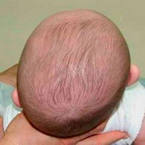
Plagiocephaly: Difference between revisions
No edit summary |
Mila Andreew (talk | contribs) No edit summary |
||
| Line 73: | Line 73: | ||
[[Category:Primary Contact]] | [[Category:Primary Contact]] | ||
[[Category:Paediatrics - Conditions]] | [[Category:Paediatrics - Conditions]] | ||
[[Category:Congenital Conditions]] | |||
Revision as of 15:25, 9 January 2023
Introduction[edit | edit source]
Deformational or positional plagiocephaly describes when a baby’s head becomes misshapen or flattened. This occurs because babies are born with soft skull bones and the junctions (sutures) between the bones are not fused. As a result, the baby’s head will sometimes become misshapen due to:
- their position in the uterus during pregnancy
- movement through the birth canal
- lying in the same position for a long time.
Positional plagiocephaly does not cause brain damage and is easily treated[1].
Clinically Relevant Anatomy[edit | edit source]
The 2 minute video below explains the skull and fontanels of a new born.
The skull covers and protects the brain and consists of several bony plates connected together by fibrous material called sutures. Sutures allow movement of the bones necessary to accommodate brain growth and allow moulding of the head during birth [3] and as a result the infant skull is vulnerable to deformation.
Mechanism of Injury / Pathological Process[edit | edit source]
Positional plagiocephaly is caused by pressure on the developing infant skull from an external force. This can occur in the womb, but more commonly develops post-natally.
Whilst practices may be different in other countries, in the UK many babies may spend significant amounts of time on their backs, either in their cot, in a car seat or in a buggy. The external forces from these firm surfaces can cause positional plagiocephaly. However it is still recommended to put babies on their backs to sleep as the importance of a reduced SIDS risk outweighs any potential dangers due to positional plagiocephaly [4].
Congenital Muscular Torticollis can also co-exist with positional plagiocephaly in as many as 30% of cases [5]. This is when a tight sternocleidomastoid muscle causes a restriction in cervical range of movement and predisposes one side of the posterior occiput to flattening.
Clinical Presentation[edit | edit source]
It is quite common for a newborn baby to have an unusually shaped head. This can be either related to their position in the uterus during pregnancy, or caused by moulding (changing shape) during labour, including changes caused by instruments used during delivery. Depending on the cause of the unusual shape, most babies' heads should go back to a normal shape within about six weeks after birth.[6]
Diagnostic Procedures[edit | edit source]
Positional plagiocephaly is diagnosed from the child's history and clinical presentation and does not usually require any imaging, however a skull x-ray may be required to rule out craniosytosis [7], which is premature fusing of the skull sutures.
Outcome Measures[edit | edit source]
As diagnosis is largely based on observation, it is helpful to record observations from different views. This can be supplemented with photography. Clinically, where no equipment is available it may be useful for parents/ carers to take photographs periodically to identify change.
Management / Interventions[edit | edit source]
It’s common for a new baby to have a flat spot on their head and in most cases this will correct itself, usually by the time the baby is sitting independently. Sometimes a baby's head does not return to a normal shape, or they may have developed a flattened spot at the back or side of their head. Sometimes a flat spot develops when a baby has limited neck movement and prefers resting their head in one particular position.[6]
You can reduce the effects of plagiocephaly by varying the position of your baby’s head and ensuring they don’t rest for long periods on the flat spot:
- Sleep time: alternate your baby’s head position from the right to the left while they sleep. It’s still important to ensure your baby sleeps on its back to help prevent Sudden Infant Death Syndrome. See the safe sleeping guidelines – Queensland Health
- Play time: Place your baby on its tummy or side during waking hours and during play time.
- Carrying and holding positions: vary how you hold or carry your baby with slings and during cuddles (over your shoulder or over your arm while they are on their tummy or side).
Plagiocephaly usually improves with time and there is no evidence to support the use of cranial remodelling helmets for babies who are healthy and developing normally.[1]
Physiotherapy[edit | edit source]
If treatment is necessary the baby attends a specialist clinic (eg a paediatrician, plastic surgeon, physiotherapist and orthotist).
The most common treatment is provided by the physiotherapist who will encourage active movement, and teach parents how to position their baby and do exercises with them to help improve the head shape.
A very small number of babies with plagiocephaly (less than one in 10) have a severe and persistent deformity, and they may need to be treated with helmet therapy.
The following video outlines the concept of Tummy Time
Differential Diagnosis[edit | edit source]
Congenital Muscular Torticollis (CMT)[edit | edit source]
A shortened sternocleidomastoid muscle can cause flattening of the occiput on the contralateral side e.g. a child with a left sided CMT presents with a right sided positional plagiocephaly. Active and passive neck movements should be checked to rule out CMT as the cause of the plagiocephaly. Early physiotherapy input is required to restore the range of movement in the neck and improve the plagiocephaly [9].
Unilateral Lambdoid Synostosis[edit | edit source]
This is rare, but caused by the premature fusion of one lambdoid suture. It is identified by retraction of the ipsilateral ear and forehead and a trapezoid shape of the head when viewed fromabove [9].
Unilateral Coronal Synostosis[edit | edit source]
Premature fusion of a coronal suture resulting in forehead assymetry and diagnosed by examining orbital symmetry. Looking from the front the ipsilateral will be higher and wider and when viewed from above the ipsilateral eyeball to the side of forehead flattening protrudes [9].
References[edit | edit source]
- ↑ 1.0 1.1 Childerens health qld gov. Plagiocephaly Available:https://www.childrens.health.qld.gov.au/fact-sheet-plagiocephaly/ (accessed 8.10.2021)
- ↑ Dr. J. Baby Skull. Available from https://www.youtube.com/watch?v=G1XhXvrWmAE&t= [Accessed 14/6/2018]
- ↑ University of Rochester Medical Centre. Anatomy of the newborn skull. https://www.urmc.rochester.edu/encyclopedia/content.aspx?contenttypeid=90&contentid=p01840 (accessed 13 June 2018).
- ↑ Great Ormond Street Hospital for Children. Positional Plagiocephaly. https://www.gosh.nhs.uk/conditions-and-treatments/conditions-we-treat-index-page-group/positional-plagiocephaly (Accessed 14 June 2018)
- ↑ Ellenbogen RG, Abdulrauf SI, Sekhar LN Principles of Neurological Surgery. Philedelphia: Elsevier, 2018.
- ↑ 6.0 6.1 RCHM Plagiocephaly – misshapen head Available:https://www.rch.org.au/kidsinfo/fact_sheets/Plagiocephaly_misshapen_head/ (accessed 8.10.2021)
- ↑ Reece A, Cohn A. Clinical Cases in Pediatrics: A trainee handbook. London: JP Medical Ltd, 2014.
- ↑ Pathways. Five essential Tummy Time moves. Available from: https://www.youtube.com/watch?v=M3rCtW9DMD4 [accessed 14/6/2018]
- ↑ 9.0 9.1 9.2 BC Children's Hospital. A Clinician's Guide to Positional Plagiocephalyhttp://www.bcchildrens.ca/neurosciences-site/Documents/BCCH034PlagiocephalyCliniciansGuideWeb1.pdf (accessed 14 June 2018)


