Pectineus Muscle
Original Editor - Your name will be added here if you created the original content for this page.
Top Contributors - Abdallah Ahmed Mohamed, Lucinda hampton, Kim Jackson and Vidya Acharya
Description[edit | edit source]
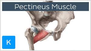
{from the Latin word pecten, meaning comb} is a flat, quadrangular muscle, situated at the anterior part of the upper and medial (inner) aspect of the thigh. The pectineus muscle is the most anterior adductor of the hip[1].
It can be classified in the medial compartment of thigh(when the function is emphasized) or the anterior compartment of thigh [2] (when the nerve is emphasized).
Origin[edit | edit source]
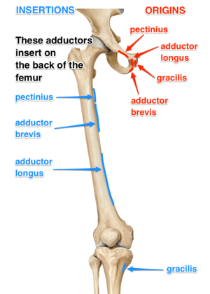
It has the most superior attachment of all the thigh adductors, originating from the pectineal line of pubis on the superior pubic ramus. The muscle then slides over the superior margin of superior pubic ramus and courses posterolaterally down the thigh, sometimes being partially divided into a larger anterior (superficial) layer and smaller posterior (deep) layer. The layers adhere to each other but are innervated by different nerves[3].
Insetion[edit | edit source]
Pectineus muscle inserts into the posterior surface of femur, along the pectineal line and proximal part of linea aspera[3].
Nerve supply[edit | edit source]
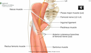
Pectineus is predominately innervated by the femoral nerve (L2, L3)]. However, in some people pectineus may receive innervation from two separate nerves of the lumber plexus[4].
This composite innervation reflects the dual compartmentalisation of pectineus into both the anterior and medial compartments of the thigh. In these cases the anterior part of the muscle sits is innervated by the femoral nerve (L2, L3), a feature of muscles of the anterior thigh. While the posterior, smaller part of the muscle is supplied by a branch of obturator nerve (L2, L3), the accessory obturator nerve[4].
Blood supply[edit | edit source]
The superficial part of the muscle is supplied by the medial circumflex femoral artery, a branch of the femoral artery. Deep portion of the muscle is vascularised by the anterior branch of obturator artery, itself a branch of the internal iliac artery.
Relation[edit | edit source]
The muscle lies in the frontal plane and medially to, adductor longus. While laterally, it is related to the psoas major muscle and the medial circumflex femoral artery and vein.
The anterior surface of pectineus forms the medial part of the floor of femoral triangle together with adductor longus.
This surface of pectineus is covered with the deep layer of fascia lata that separates it from the femoral artery, femoral vein and great saphenous vein that course through the femoral triangle.
Posterior to pectineus are the adductor magnus, adductor brevis and obturator externus muscles, and the anterior branch of obturator nerve.[3][4]
Resources[edit | edit source]
- bulleted list
- x
or
- numbered list
- x
References[edit | edit source]
- ↑ Mosby's Medical, Nursing & Allied Health Dictionary, Fourth Edition, Mosby-Year Book Inc., 1994, p. 1177
- ↑ Ellis, Harold; Susan Standring; Gray, Henry David (2005). Gray's anatomy: the anatomical basis of clinical practice. St. Louis, Mo: Elsevier Churchill Livingstone. p. 518.
- ↑ 3.0 3.1 3.2 Moore, K. L., Dalley, A. F., & Agur, A. M. R. (2014). .Clinically Oriented Anatomy (7th ed).
- ↑ 4.0 4.1 4.2 Standring, S. (2016). aGray's Antomy (41tst ed.). Edinburgh: Elsevier Churchill Livingstone.


