Multiligament Injured Knee Dislocation: Difference between revisions
No edit summary |
No edit summary |
||
| Line 114: | Line 114: | ||
== Outcome Measures == | == Outcome Measures == | ||
Recent prospective studies declare that patients who achieve a good Lysholm score, could perform activities on a regular basis and could perform hop tests comparable to the other knee.<br> | Recent prospective studies declare that patients who achieve a good Lysholm score, could perform activities on a regular basis and could perform hop tests comparable to the other knee.<ref name="1">N.R. Howells, et al.; „Acute knee dislocation: An evidence based approach to the management of the multiligament injured knee”; Injury; 2010. Level of evidence 3A</ref><br> | ||
== Examination == | == Examination == | ||
Revision as of 19:08, 1 January 2014
Original Editors - Caro De Koninck
Top Contributors - Estelle Hovaere, Leana Louw, Caro De Koninck, Admin, Kim Jackson, 127.0.0.1, Evan Thomas, Daphne Jackson, WikiSysop, Naomi O'Reilly and Wanda van Niekerk
Search Strategy[edit | edit source]
Key-words: knee dislocation, knee injury, total knee dislocation, multiligament knee dislocation
Definition/Description[edit | edit source]
A knee dislocation describes the complete disruption of the tibiofemoral articulation. This results in a multiligament injury which is described as a rupture in two of the four major knee ligament structures: anterior cruciate ligament (ACL), posterior cruciate ligament (PCL), posterolateral corner (lateral collateral (LCL) , popliteus and popliteofibular ligament) (PCL) and medial collateral ligament (MCL).Cite error: Invalid <ref> tag; name cannot be a simple integer. Use a descriptive title (N.R. Howells, 2010, 3A)
Knee dislocations occur in 5 main types: anterior, posterior, medial, lateral and rotary. The anterior dislocations represent more than 70% of knee dislocations. Rotary dislocations can further be divided into anteromedial, anterolateral, posteromedial and posterolateral injuries.[1] (Henrichs, 2004, A1)
Clinically Relevant Anatomy[edit | edit source]
add text here
Epidemiology /Etiology[edit | edit source]
Knee dislocation is estimated to be less than 0,02% of all orthopedic injuries. Total knee dislocations are rare. They usually happen after major trauma, including falls, car crashes, and other high-speed injuries. Even spontaneous dislocation is possible but most of the time they are associated with obesity. Cite error: Invalid <ref> tag; name cannot be a simple integer. Use a descriptive title
It can also be a birth deformity. Congenital dislocation of the knee has an incidence of approximately 1 per 100,000 live births. 40-100% of these cases have additional musculoskeletal anomalies. [2](L. Flint, A)
Characteristics/Clinical Presentation[edit | edit source]
Acute knee dislocations are uncommon orthopaedic injuries. Because they often spontaneously reduce before initial evaluation, the true incidence is unknown. Dislocation can be suspected based on physical exam findings of joint instability/ligamentous injuries, but also based on hemarthosis and tenderness to palpation. (D. Shearer, 1A)[3] Associated meniscal, osteochondral, and neurovascular injuries are often present and can complicate management. (Rihn et al., 2004, A1)[4]
Ligamentous injuries have characteristics such as:
- Long-term joint instability
- Patient’s mobility
- Quality of life
The most common problems of knee dislocation are stiffness, secondary to arthrofibrosis or failure of a ligament repair or reconstruction. In the long term, it is reported that up to 50% of patients may develop post-traumatic Osteoarthritis
Differential Diagnosis[edit | edit source]
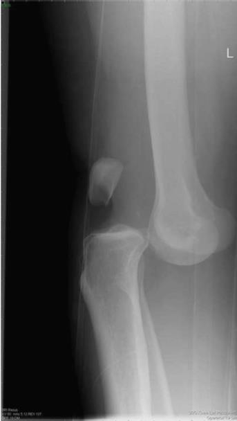
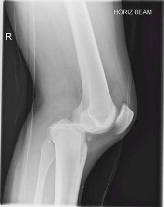
Classifications of knee dislocations have been based on either an anatomical and/or a positional scheme. The positional classification categorizes knee dislocations according to the tibial position in relation to the femur also known as the Kennedy Classification, named after the author in 1963. Five types were initially defined Cite error: Invalid <ref> tag; name cannot be a simple integer. Use a descriptive title :
- anterior (Fig. 1)
- posterior (Fig. 2)
- lateral
- medial
- rotatory
Rotatory knee dislocations were subdivided into four groups: anteromedial, anterolateral, posteromedial and posterolateral.
Although well established and useful, the positional classification system has limitations. Up to 50% of knee dislocations are spontaneously reduced before evaluation and cannot be classified with the positional classification. Therefore an anatomical system was developed, based on ligament injury with additional designations of C (arterial injury) and N (neural injury)[5].
KDI is described as —> a single cruciate tear
KDII —> bicruciate tears without collateral tears
KDIIIM —> bicruciate tears with involvement of a medial collateral ligament
(MCL) KDIIIL —> involvement of a lateral collateral ligament (LCL) and
posterolateral corner (PLC) tear
KDIV —> involves all four ligaments and KDV involves a fracture-dislocation.
In general, the higher the number, the greater the injury to the knee.
Diagnostic Procedures[edit | edit source]
Physical examination of a patient with a suspected knee dislocation should take place shortly after the injury is sustained. (Levy et al., 2010) Recognition is the most important aspect of the diagnosis. When a knee dislocation is suspected, neurovascular assessment is needed. Patients with a knee dislocation complain of severe pain and instability. Also they are unable to continue sports or activities of daily living. Pain tends to be diffuse with palpation and knee ROM's are limited. The Lachman and pivot-shift tests should be performed to test ACL integrity and the posterior drawer and posterior sag tests should be performed to test PCL integrity. Varus and Valgus stress tests should be carried out to test for MCL and LCL injury. (Henrichs, 2004, A1)[6]
All cases of suspected knee dislocation should have an ankle-brachial index performed, reserving arteriography for those with an abnormal finding. (Levy et al., 2010, A1)[7]
Outcome Measures[edit | edit source]
Recent prospective studies declare that patients who achieve a good Lysholm score, could perform activities on a regular basis and could perform hop tests comparable to the other knee.Cite error: Invalid <ref> tag; name cannot be a simple integer. Use a descriptive title
Examination[edit | edit source]
add text here related to physical examination and assessment
Medical Management
[edit | edit source]
Definitive management of acute knee dislocation remains a matter of debate; however, surgical reconstruction or repair of all ligamentous injuries likely can help in achieving the return of adequate knee function. (Rihn et al., 2004, A1)[4]
Also Henrichs (2004, A1)[6] says that surgical treatment had proven to be much more beneficial for active patients. Conservative treatment on the other hand is often chosen if the joint feels relatively stable postreduction or if the patient is older or sedentary with intact colletarel ligaments.
Physical Therapy Management
[edit | edit source]
A patient with a knee dislocation is faced with a long rehabilitation program, with return to full activity taking at least 9 to 12 months.
After conservative treatment, rehabilitation may begin immediately. The first exercises are upper and midbody exercises, along with single-leg stationary bicycling in order to maintain
cardiovascular conditioning. Quadriceps strengthening is important to prevent patellofemoral problems during rehabilitation. During early exercises, wearing a brace is important to limit ROM from 90°of flexion to 45°of extension. Light manual-resistance exercises may be performed in this range as tolerated. As the patient progresses, other resistance machines such as Biodex may be used in place of manual therapy. After 8 weeks of rehabilitation, the patient may begin doing exercises with a leg-press machine. During this face, the knee needs only minimal protection. These exercises should be followed by high-speed exercises with light resistance. Once sufficient ROM and strength have been regained, proprioceptive exercises may be integrated.
Postsurgical rehabilitation for this injury varies according to which ligaments were injured and repaired. Initially, a brace with limits of 40° of extension and 70° of flexion should be worn if a continuous passive-motion machine is used. From the start, quadriceps settings are performed in order to increase quadriceps control. In the first postoperative week it's also very important to work on full extension. This can be achieved by doing therapeutic exercises. From weeks 7 to 10, ROM therapy can begin, stressing full extension and flexion. After weeks 11 to 24, the patient should have regained full flexion and extension and strength training can begin using closed kinetic chain exercises (squats, light leg presses,..). Strength training continues through weeks 25 to 36 but becomes more advanced. After the patient has reestablished bulk and strength around the knee, proprioceptive exercises are integrated. After week 37 the patient can return to sports and heavy work, provided that he or she has passed functional tests.
(Henrichs, 2004, A1)[6]
Key Research[edit | edit source]
add links and reviews of high quality evidence here (case studies should be added on new pages using the case study template)
Resources
[edit | edit source]
Web MD, Knee pain health center. http://www.webmd.com/pain-management/knee-pain/knee-dislocation (assessed 14 april 2011)
Henrichs A. A review of knee dislocations. Journal of Athletic Training 2004;39(4): 365–369
Rihn J, Groff Y, Harner C & Cha P. The acutely dislocated knee: evaluation and management. J Am Acad Orthop Surg. 2004 Sep-Oct;12(5): 334-46.
Levy B, Peskun C, Fanelli G, Stannard J, Stuart M, MacDonald P, Marx R, Boyd J & Whelan D. Diagnosis and management of knee dislocations. Phys Sportsmed. 2010 Dec;38(4): 101-11.
Clinical Bottom Line[edit | edit source]
add text here
Recent Related Research (from Pubmed)[edit | edit source]
see tutorial on Adding PubMed Feed
Extension:RSS -- Error: Not a valid URL: Feed goes here!!|charset=UTF-8|short|max=10
References[edit | edit source]
- ↑ Henrichs A. A review of knee dislocations. Journal of Athletic Training 2004;39(4): 365–369. level of evidence: A1
- ↑ Lewis Flint et co, Trauma: contemporary principles and therapy. Grade of recommendation A
- ↑ LCDR Damon Shearer, DO; Laurie Lomasney, MD; Kenneth Pierce, MD. Dislocation of the knee: imaging findings, J Spec Oper Med. 2010 Winter;10(1):43-7. Level of evidence: A1
- ↑ 4.0 4.1 Rihn J, Groff Y, Harner C &amp;amp; Cha P. The acutely dislocated knee: evaluation and management. J Am Acad Orthop Surg. 2004 Sep-Oct;12(5): 334-46. A1
- ↑ R. Schenck Jr. MD „Classification of knee dislocations” Operative Techniques in Sports Medicine, Vol 11, No 3 (July), 2003) Level of evidence 2A
- ↑ 6.0 6.1 6.2 Cite error: Invalid
<ref>tag; no text was provided for refs namedHenrichs et al. - ↑ Levy B, Peskun C, Fanelli G, Stannard J, Stuart M, MacDonald P, Marx R, Boyd J & Whelan D. Diagnosis and management of knee dislocations. Phys Sportsmed. 2010 Dec;38(4): 101-11. A1
see adding references tutorial.


