Medical Imaging
Overview[edit | edit source]
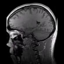
Medical imaging is home to all diagnostic and therapeutic investigations/interventions conducted in a typical radiology department. It encompasses different imaging modalities and processes to image the human body for diagnostic, treatment and follow up purposes and plays an important role in initiatives to improve public health for all population groups[1]. It includes:
X-rays (including eg plain xrays, DEXA scans, fluoroscopy)
- Ultrasound (US)
- Computed tomography (CT)
- nuclear medicine: often cross-sectional radiotracer scanning e.g. PET is considered a separate modality from 'traditional' scintigraphy e.g. bone scans
- Hybrid modalities[2]
Medical imaging, especially X-ray based examinations and ultrasonography, is crucial in a variety of medical setting and at all major levels of health care. In public health and preventive medicine as well as in both curative and palliative care, effective decisions depend on correct diagnoses. Though medical/clinical judgment may be sufficient prior to treatment of many conditions, the use of diagnostic imaging services is paramount in confirming, correctly assessing and documenting courses of many diseases as well as in assessing responses to treatment
Imaging is a useful resource for many conditions and is an invaluable tool for physical therapists when used appropriately.
It is important to know when imaging is appropriate, as unnecessary imaging will squander financial resources and increase potential for premature surgery.
Nuclear Medicine[edit | edit source]
Nuclear medicine in vivo is the practice of utilising small amounts of radioactive substances (unsealed radioactive sources) to diagnose, monitor and treat disease. The utilisation of radiopharmaceuticals offers a unique perspective on both disease and cancer treatment[4].
Included in Nuclear Medicine are:
Bone scan is an imaging technique that uses a radioactive compound to identify areas of healing within the bone. Bone scans work by drawing blood from the patient and tagging it with a bone seeking radiopharmaceutical. This radioactive compound emits gamma radiation. The blood is then returned to the patient intravenously. As the body begins its metabolic activity at the site of the injury, the blood tagged by the radioactive compound is absorbed at the bone and the gamma radiation at the site of the injury can be detected with an external gamma camera. A bone scan can be beneficial in determining injury to the bone within the first 24-48 hours of injury or when the displacement is too small to be detected by an x-ray or CT scan.[5].[6]
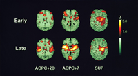
2. Positron Emission Tomography (PET) is primarily used to detect diseases of the brain and heart. Similarly to nuclear medicine, a short-lived isotope, such as 18F, is incorporated into a substance used by the body such as glucose which is absorbed by the tumor of interest. PET scans are often viewed alongside computed tomography scans, which can be performed on the same equipment without moving the patient. This allows the tumors detected by the PET scan to be viewed next to the rest of the patient's anatomy detected by the CT scan.
3. Single Photon Emission Computed Tomography (SPECT) is a widely used imaging technique in nuclear medicine for the visualization of organs, such as the bones, heart and brain, as well as for the detection of tumors.[7] Because of its capability to visualize and quantify changes in the cerebral blood flow and neurotransmitter system, it has important use in the differential diagnosis of neurological and psychiatric diseases. [8]
Optoacoustic Imaging[edit | edit source]
Also known as Photoacoustic Imaging, is an upcoming biomedical imaging modality availing the benefits of optical resolution and acoustic depth of penetration. With its capacity to offer structural, functional, molecular and kinetic information making use of either endogenous contrast agents like hemoglobin, lipid, melanin and water or a variety of exogenous contrast agents or both, Optoacoustic imaging has demonstrated promising potential in a wide range of preclinical and clinical applications.[9]
Clinical applications of optoacoustic imaging include:[9]
- Breast imaging
- Dermatologic Imaging
- Pilosebaceous units
- Skin cancer
- Inflammatory skin diseases
- Vascular Imaging
- Cutaneous miscrovasculature
- Vascular Dysfunction
- Wound Imaging
- Carotid Vessel Imaging
- Musculoskeletal Imaging
- Gastrointestinal Imaging
- Adipose Tissue Imaging
Recent studies showed the potential use of optoacoustic imaging in the assessment, diagnosis and monitoring of treatment in patients with inflammatory arthritis[10] as well limb and muscle ischemia.[11]
Health Care Team[edit | edit source]
Imaging for medical purposes involves a team which includes the service of radiologists, radiographers (X-ray technologists), sonographers (ultrasound technologists), medical physicists, nurses, biomedical engineers, and other support staff working together to optimize the wellbeing of patients, one at a time. Appropriate use of medical imaging requires a multidisciplinary approach.[1]
Diagnostic Imaging for Body Regions[edit | edit source]
- Diagnostic Imaging of the Hip for the Physical Therapist
- Diagnostic Imaging of the Knee for the Physical Therapist
- Diagnostic Imaging of the Ankle and Foot for the Physical Therapist
- Diagnostic Imaging of the Shoulder
References[edit | edit source]
- ↑ 1.0 1.1 WHO Diagnostic Imaging Available from: https://www.who.int/diagnostic_imaging/en/(accessed 7.4.2021)
- ↑ Radiopedia Modalities Available from:https://radiopaedia.org/articles/modality?lang=us (accessed 7.4.2021)
- ↑ The Audiopedia What is Medical Imaging? What does Medical Imaging mean? Medical Imaging meaning & explanation Available from: https://www.youtube.com/watch?v=Dm9iaq8uMkI (last accessed 1.10.19)
- ↑ Radiopedia Nuclear medicine Available from: https://radiopaedia.org/articles/nuclear-medicine?lang=gb (accessed 7.4.2021)
- ↑ Swain J, Bush K. Diagnostic Imaging for Physical Therapists. St. Louis: Saunders Elsevier; 2009
- ↑ College A. ACR Practice Guideline For The Performance Of Adult and Pediatric Skeletal Scintigraphy ( Bone Scan ). North. 2007:1-5.
- ↑ Warwick, J.M. Imaging of Brain Function Using SPECT. Metab Brain Dis 19, 113–123 (2004) obtained from https://link.springer.com/article/10.1023/B:MEBR.0000027422.48744.a3 doi:10.1023/B:MEBR.0000027422.48744.a3
- ↑ Andrew B. Newberg, Abass Alavi, Single Photon Emission Computed Tomography☆, Reference Module in Neuroscience and Biobehavioral Psychology, Elsevier, 2017, ISBN 9780128093245, obtained from https://www.sciencedirect.com/science/article/pii/B9780128093245024871 doi: 10.1016/B978-0-12-809324-5.02487-1
- ↑ 9.0 9.1 Amalina Binte Ebrahim Attia, Ghayathri Balasundaram, Mohesh Moothanchery, U.S. Dinish, Renzhe Bi, Vasilis Ntziachristos, Malini Olivo, A review of clinical photoacoustic imaging: Current and future trends, Photoacoustics, Volume 16, 2019, 100144, ISSN 2213-5979, obtained from https://www.sciencedirect.com/science/article/pii/S2213597919300679 doi: 10.1016/j.pacs.2019.100144
- ↑ Jo J, Tian C, Xu G, et al. Photoacoustic tomography for human musculoskeletal imaging and inflammatory arthritis detection. Photoacoustics. 2018;12:82–89. Published 2018 Jul 27 obtained from https://www.ncbi.nlm.nih.gov/pmc/articles/PMC6306364/ doi:10.1016/j.pacs.2018.07.004
- ↑ Chen L, Ma H, Liu H, et al. Quantitative photoacoustic imaging for early detection of muscle ischemia injury. Am J Transl Res. 2017;9(5):2255–2265. Published 2017 May 15 obtained from https://www.ncbi.nlm.nih.gov/pmc/articles/PMC5446508/


