Kinematics of the Cervical Spine
The cervical spine is the most mobile region of the spine and enables the head with its sensory organs of sight, hearing and smell to be oriented independently from the trunk. The kinematics of the cervical spine are complex - seven vertebrae as well as articulations with the occiput and upper thoracic spine, each with different axes of movement which can combine in a multitude of ways. As a result, an intended orientation of the head can be achieved in multiple ways.
Movements of the cervical spine are 1) constrained by the anatomy of motion segments and longer connective structures that span multiple segments and 2) controlled by the neuromuscular system (insert link) both of which are discussed elsewhere. This section will consider active movement (AROM) of the neck and of the individual motion segments that make up the neck from a kinematics perspective - that is the movements will be discussed without considering how the movements are constrained or produced.
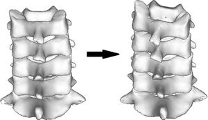
Although this section will consider the cervical spine, it is important to recognize that the neck is connected to the rest of the body. Movement of the head includes movement of the thoracis as well as the cervical spine (Figure 1)
Cardinal Directions[edit | edit source]
Positions and movements of the body are generally described in relation to three axes of movement and three cardinal planes (link to Cardinal Planes and Axes of Movement). In the spine, and particularly the cervical spine, movements do not occur simply in one plane or around one axis, but rather include translational movements as well as coupled (also referred to as conjoint or combined movements) movements around other axes [1] .
Active Movements[edit | edit source]
Active cervical rotation usually results in a small amount of coupled lateral flexion and flexion/extension (Figure 2) with the coupled movements of lateral flexion are usually greater than those in flexion/extension. Different studies have found that the average lateral flexion is either ipsilateral [2] or the contralateral [1] to the direction of rotation.
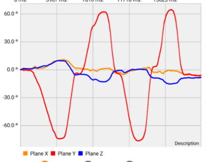
When considering the observed coupled movements Salem, et. al. (2) stated:
Mean coupled lateral flexion was −2.3 ± 10.7° occurring in the same direction as the rotation. However, the standard deviation is important, indicating that several subjects performed a lateral flexion in the opposite direction to rotation (5 of 20 subjects).
Not only is this coupling not consistent between individuals, but it also varies with repeated movements for a given individual. How someone is instructed to do move may also change the pattern. For example, “turn your chin towards your left shoulder” is likely to result in a different pattern of movement than “turn to see as far as you can behind your left shoulder”. Some translation of the head also occurs. Perhaps most notably there is usually a translation towards the side of rotation.
Segmental Movement[edit | edit source]
Contributions of individual motion segments of the cervical spine to full range AROM[edit | edit source]
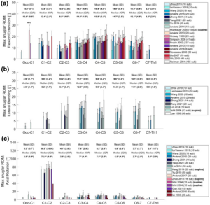
The amount of movement in of motion segments (movement between individual pairs of vertebrae) varies between levels as shown below. Note that these ranges are for full axis movements from the limit in one direction to the limit in the opposite direction.
The range of rotation of C1-C2 makes up approximately half of the total AROM range. The other motion segments from C2 – C7 generally have less than 11 degrees of rotation with even less at O – C1. The segmental movements in flexion/extension are fairly similar across the region (10.0 to 16.4°) except for less movement occurring in C7-T1. Lateral flexion is also fairly consistent at around 10 degrees per level from C2 to C7 but with less movement between the occiput and C2.
For one-sided movements, the mean lateral flexion between C2 and C7 is about five degrees in each direction while the mean rotation from C3 to C7 ranges from two to four degrees.
An aside[edit | edit source]
The descriptions of the averages above for both coupled and segmental movement patterns however have limitations as suggested by the English polymath and statistician Frances Galton (1822 – 1911) who so eloquently stated:
“It is difficult to understand why statisticians commonly limit their inquiries to averages, and do not revel in more comprehensive views. Their souls seem as dull to the charm of variety as that of the native of one of our flat English counties, whose retrospect of Switzerland was that, if its mountains could be thrown into its lakes, two nuisances would be got rid of at once.”
Axes of Movement[edit | edit source]
The structure of the facets and discs determine the movements that are possible between each pair of vertebrae. During AROM, the facets of the cervical spine mostly remain in contact [3]. There are some exceptions to this. For example there is often some separation of the facet surfaces in the upper cervical spine during rotation that enables the head to remain level.
Because of these structures, the segmental movements for C1 – T1 do not occur along the three cardinal planes but can be better described as occurring around two perpendicular axes. [4] Flexion/Extension occur around one transverse axis which passes through ne vertebral body of the lower vertebra in the motion segment. A second axis is perpendicular to the first and is approximately normal to the plane of the facets. Just as the plane of the facets is different for different levels, so too is the orientation of the second axis. This second axis is more vertical in the upper cervical spine and moves closer to an AP direction for the lower motion segments. Importantly, the axes are not fixed, and the instantaneous centres of rotation (the location of the axis of movement) move somewhat during active movement.
Coupled Movements – Segmental[edit | edit source]
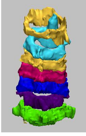
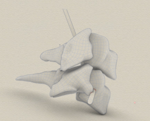
There is usually an ipsilateral lateral flexion in the lower cervical motion segments during active rotation. An image of the segments of the lower cervical spine at the end of range of rotation is shown below. This image shows the most common pattern that occurs at the limit of rotation. Each of the motion segments from C3 to C7 have the facets moving down on the right and up on the left to create a position of C3 which is both rotated and laterally flexed to the right. Image from (4) doi: https://doi.org/10.1371/journal.pone.0215357.g005 creative commons license
As well as being rotated in this position, C3 is tilted (laterally flexed) to the ipsilateral side. The upper cervical spine therefore needs to do some adjusting to keep the head level while facing to the right. These movement strategies include translations as well coupled movements around other than the primary axis of the upper cervical spine.
Segmental movement[edit | edit source]
The amount of movement in of motion segments (movement between individual pairs of vertebrae) varies between levels as shown in the figure below. The mean values from several studies as shown here are fairly consistent. As will be described later, it is important to recognize that there remains a high level of variation between individuals.
- ↑ 1.0 1.1 Kang J, Chen G, Zhai X, He X. In vivo three-dimensional kinematics of the cervical spine during maximal active head rotation. PLoS One. 2019;14(4):e0215357.
- ↑ Salem W, Lenders C, Mathieu J, Hermanus N, Klein P. In vivo three-dimensional kinematics of the cervical spine during maximal axial rotation. Manual therapy. 2013.
- ↑ Malmstrom EM, Karlberg M, Fransson PA, Melander A, Magnusson M. Primary and Coupled Cervical Movements: The Effect of Age, Gender, and Body Mass Index. A 3-Dimensional Movement Analysis of a Population Without Symptoms of Neck Disorders. Spine. 2006;31(2):E44-E50.
- ↑ Bogduk N, Mercer S. Biomechanics of the cervical spine. I: Normal kinematics. Clin Biomech. 2000;15(9):633-48
