Kinematics of the Cervical Spine
The cervical spine is the most mobile region of the spine and enables the head with its sensory organs of sight, hearing and smell to be oriented independently from the trunk. The kinematics of the cervical spine are complex - seven vertebrae as well as articulations with the occiput and upper thoracic spine, each with different axes of movement which can combine in a multitude of ways. As a result, an intended orientation of the head can be achieved in multiple ways.
Movements of the cervical spine are 1) constrained by the anatomy of motion segments and longer connective structures that span multiple segments and 2) controlled by the neuromuscular system (insert link) both of which are discussed elsewhere. This section will consider active movement (AROM) of the neck and of the individual motion segments that make up the neck from a kinematics perspective - that is the movements will be discussed without considering how the movements are constrained or produced.
Although this section will consider the cervical spine, it is important to recognize that the neck is connected to the rest of the body. Movement of the head includes movement of the thoracis as well as the cervical spine (Figure 1)
Cardinal Directions[edit | edit source]
Positions and movements of the body are generally described in relation to three axes of movement and three cardinal planes (link to Cardinal Planes and Axes of Movement). In the spine, and particularly the cervical spine, movements do not occur simply in one plane or around one axis, but rather include translational movements as well as coupled (also referred to as conjoint or combined movements) movements around other axes [1] .
Active Movements[edit | edit source]
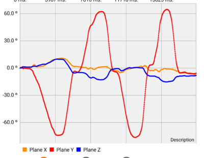
Active cervical rotation usually results in a small amount of coupled lateral flexion and flexion/extension (Figure 2) with the coupled movements of lateral flexion are usually greater than those in flexion/extension. Different studies have found that the average lateral flexion is either ipsilateral [2] or the contralateral [1] to the direction of rotation.

Figure 1. Active head rotation of the author recorded using Goniometro Advance app on an Android phone. Red is Rotation, Orange flexion/extension, and blue Lateral flexion. Vertical lines indicate the variation of these coupled movements (mean +/- 1.96*SD) of a group of participants aged in their 20’s of values from Salem et. al. 2012
When considering the observed coupled movements Salem, et. al. (2) stated:
Mean coupled lateral flexion was −2.3 ± 10.7° occurring in the same direction as the rotation. However, the standard deviation is important, indicating that several subjects performed a lateral flexion in the opposite direction to rotation (5 of 20 subjects).
Not only is this coupling not consistent between individuals, but it also varies with repeated movements for a given individual. How someone is instructed to do move may also change the pattern. For example, “turn your chin towards your left shoulder” is likely to result in a different pattern of movement than “turn to see as far as you can behind your left shoulder”. As well as coupled movements some translation also occurs with movements of the cervical spine.
- ↑ 1.0 1.1 Kang J, Chen G, Zhai X, He X. In vivo three-dimensional kinematics of the cervical spine during maximal active head rotation. PLoS One. 2019;14(4):e0215357.
- ↑ Salem W, Lenders C, Mathieu J, Hermanus N, Klein P. In vivo three-dimensional kinematics of the cervical spine during maximal axial rotation. Manual therapy. 2013.


