Kemp Test: Difference between revisions
No edit summary |
No edit summary |
||
| (10 intermediate revisions by 3 users not shown) | |||
| Line 4: | Line 4: | ||
'''Top Contributors''' - {{Special:Contributors/{{FULLPAGENAME}}}} | '''Top Contributors''' - {{Special:Contributors/{{FULLPAGENAME}}}} | ||
</div> | </div> | ||
== Description == | == Description == | ||
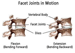
[[File:Facet-joints.png|alt=|right|frameless]]The Kemp test (also known as the quadrant test and extension-rotation test) is a provocative test useful for diagnosing pain related to [[Facet Joint Syndrome|facet joint pathology]], e.g. [[osteoarthritis]]. | |||
The | # The client performs combined extension and rotation of the spine (used for the [[Cervical Anatomy|cervical spine]] or the [[Lumbar|lumbar spine]]). | ||
# The test is considered positive when the patient reports [[Pain Assessment|pain]], numbness or tingling in the area concerned.<ref>Radiopedia Kemp Test Available:https://radiopaedia.org/articles/kemp-test?lang=gb (accessed 26.9.2022)</ref> | |||
[[ | |||
== Purpose == | == Purpose == | ||
The purpose of this test is to assess lumbar spine facet | The purpose of this test is to assess lumbar or cervical spine [[Facet Joints|facet joint]] pain. It uses the patient’s trunk both as a lever to induce tension and as a compressive force. This test is used in differentiation and diagnosis of a lumbar posterior [http://www.physio-pedia.com/index.php/Facet_Joint_Syndrome facet syndrome], though it is nonspecific.<ref name="p4">Craig E. Morris; Low back syndromes; McGraw-hill professional; 2005 (Level of evidence: D)</ref> It is a provocation test to detect pain. Local pain suggest a facet cause, whilst radiating pain into the leg/arm is more suggestive of nerve root irritation. Especially if the pain is below the knee/elbow.<ref name="p5">Souza TA. [https://books.google.com/books?hl=en&lr=&id=g8FEXRYkN2wC&oi=fnd&pg=PR1&dq=Thomas+A.+Souza%3B+differential+diagnosis+and+management+for+the+chiropractor%3B+Jones+%26+Bartlett+Learning%3B+2008+(Level+of+evidence:+D)&ots=WMksJjptOD&sig=s5MUc8GwPmwRKLYjbwl-S6 Differential diagnosis and management for the chiropractor: protocols and algorithms]. Jones & Bartlett Publishers; 2009 Oct 7.</ref> | ||
== Technique == | == Technique == | ||
The | The Kemp Test test may be performed with the patient either in the seated or standing position. | ||
Standing position: <br> 1. Patient is standing before the therapist.<br> 2. The therapist fixes the opposite ilium from the side being tested with one hand.<br> 3. The other hand grabs the shoulder from the patient and leads the patient to extension, ipsilateral side bending and | '''Standing position''': <br> 1. Patient is standing before the therapist.<br> 2. The therapist fixes the opposite ilium from the side being tested with one hand.<br> 3. The other hand grabs the shoulder from the patient and leads the patient to extension, ipsilateral side bending and ipsilateral rotation (3D extension movement).<br> 4. Hold this position for three seconds. | ||
{{#ev:youtube|tBVhHpxF3ZQ|300}} <ref>Rocky Bains. Kemp's Test. Available from: https://www.youtube.com/watch?v=tBVhHpxF3ZQ [last accessed 22/10/2020]</ref> | {{#ev:youtube|tBVhHpxF3ZQ|300}} <ref>Rocky Bains. Kemp's Test. Available from: https://www.youtube.com/watch?v=tBVhHpxF3ZQ [last accessed 22/10/2020]</ref> | ||
Seated position:<br> 1. The patient seated with arms crossed over the chest.<br> 2. One hand of the therapist stabilize the patient’s lumbosacral region on the side to be tested.<br> 3. The other arm controls the patient’s upper body movement.<br> 4. The patient is passively directed into flexion, rotation, lateral flexion, and finally extension.<br> 5. Depending on the patient’s response, axial compression may be applied in the fully extended and rotated position to increase stress on the posterior joints. <br> | '''Seated position''':<br> 1. The patient seated with arms crossed over the chest.<br> 2. One hand of the therapist stabilize the patient’s lumbosacral region on the side to be tested.<br> 3. The other arm controls the patient’s upper body movement.<br> 4. The patient is passively directed into flexion, rotation, lateral flexion, and finally extension.<br> 5. Depending on the patient’s response, axial compression may be applied in the fully extended and rotated position to increase stress on the posterior joints. <br> | ||
The test is positive when the patient reports pain, numbness or tingling in the area of the back or lower extremities. The pain is located on the side being tested.<ref name="p1">Steve Jensen; back pain – clinical assessment; Australian Family Physician Vol. 33, No. 6, June 2004 (level of evidence:D)</ref><ref name="p3">Lyle MA, Manes S, McGuinness M, Ziaei S, Iversen MD. [https://academic.oup.com/ptj/article-abstract/85/2/120/2804972 Relationship of physical examination findings and self-reported symptom severity and physical function in patients with degenerative lumbar conditions.] Physical therapy. 2005 Feb 1;85(2):120-33.</ref><ref name="p4" /><span style="font-size: 11px;"> </span>Local pain suggests a facet cause, while radiating pain into the leg is more suggestive of [http://www.physio-pedia.com/index.php/Lumbar_Radiculopathy#top nerve root irritation]. Especially if the pain is below the knee.<sup><ref name="p5" /></sup><br>The seated position is more preferable because the therapist has more control over the patient’s positioning and there is less muscle activation.<sup><ref name="p4" /><ref name="p5" /></sup><sup></sup> | The test is positive when the patient reports pain, numbness or tingling in the area of the back or lower extremities. The pain is located on the side being tested.<ref name="p1">Steve Jensen; back pain – clinical assessment; Australian Family Physician Vol. 33, No. 6, June 2004 (level of evidence:D)</ref><ref name="p3">Lyle MA, Manes S, McGuinness M, Ziaei S, Iversen MD. [https://academic.oup.com/ptj/article-abstract/85/2/120/2804972 Relationship of physical examination findings and self-reported symptom severity and physical function in patients with degenerative lumbar conditions.] Physical therapy. 2005 Feb 1;85(2):120-33.</ref><ref name="p4" /><span style="font-size: 11px;"> </span>Local pain suggests a facet cause, while radiating pain into the leg is more suggestive of [http://www.physio-pedia.com/index.php/Lumbar_Radiculopathy#top nerve root irritation]. Especially if the pain is below the knee.<sup><ref name="p5" /></sup><br>The seated position is more preferable because the therapist has more control over the patient’s positioning and there is less muscle activation.<sup><ref name="p4" /><ref name="p5" /></sup><sup></sup> | ||
{{#ev:youtube|4wIFZmxiSTg|300}}<ref>Palmer Health Sciences Library. Kemp's Test. Available from: https://www.youtube.com/watch?v=75bVhJ-sBcI [last accessed 22/10/2020]</ref> | {{#ev:youtube|4wIFZmxiSTg|300}}<ref>Palmer Health Sciences Library. Kemp's Test. Available from: https://www.youtube.com/watch?v=75bVhJ-sBcI [last accessed 22/10/2020]</ref> | ||
The below video is of the Kemp test in the cervical spine (similar to [[Spurling's Test]]). | |||
{{#ev:youtube|v=jNdBq3eR-eY|300}} <ref>Massage nerd. Orthopedic Test - C / SPINE KEMP'S TEST. Available from: https://www.youtube.com/watch?v=jNdBq3eR-eY [last accessed 26.9.2022]</ref> | |||
== Evidence == | == Evidence == | ||
Kemp’s test is the most used | Kemp’s test is the most used diagnostic procedure for lumbar pain but has a poor diagnostic accuracy with a sensitivity of 50-70%<ref name="p3" /> and specificity of 67.3%<ref>Manchikanti L, Pampati V, Fellows B, Baha AG. [https://www.painphysicianjournal.com/current/pdf?article=MzMw&journal=3 The inability of the clinical picture to characterize pain from facet joints]. Pain Physician. 2000 Apr;3(2):158-66.</ref>. | ||
== Resources == | == Resources == | ||
Latest revision as of 23:29, 18 December 2023
Original Editor - Rachael Lowe
Top Contributors - Aminat Abolade, Admin, Rachael Lowe, Kim Jackson, Lucinda hampton, WikiSysop, Wanda van Niekerk, Jess Bell, 127.0.0.1, Tony Lowe, Roel De Groef, Aarti Sareen, Oyemi Sillo and Kai A. Sigel
Description[edit | edit source]
The Kemp test (also known as the quadrant test and extension-rotation test) is a provocative test useful for diagnosing pain related to facet joint pathology, e.g. osteoarthritis.
- The client performs combined extension and rotation of the spine (used for the cervical spine or the lumbar spine).
- The test is considered positive when the patient reports pain, numbness or tingling in the area concerned.[1]
Purpose[edit | edit source]
The purpose of this test is to assess lumbar or cervical spine facet joint pain. It uses the patient’s trunk both as a lever to induce tension and as a compressive force. This test is used in differentiation and diagnosis of a lumbar posterior facet syndrome, though it is nonspecific.[2] It is a provocation test to detect pain. Local pain suggest a facet cause, whilst radiating pain into the leg/arm is more suggestive of nerve root irritation. Especially if the pain is below the knee/elbow.[3]
Technique[edit | edit source]
The Kemp Test test may be performed with the patient either in the seated or standing position.
Standing position:
1. Patient is standing before the therapist.
2. The therapist fixes the opposite ilium from the side being tested with one hand.
3. The other hand grabs the shoulder from the patient and leads the patient to extension, ipsilateral side bending and ipsilateral rotation (3D extension movement).
4. Hold this position for three seconds.
Seated position:
1. The patient seated with arms crossed over the chest.
2. One hand of the therapist stabilize the patient’s lumbosacral region on the side to be tested.
3. The other arm controls the patient’s upper body movement.
4. The patient is passively directed into flexion, rotation, lateral flexion, and finally extension.
5. Depending on the patient’s response, axial compression may be applied in the fully extended and rotated position to increase stress on the posterior joints.
The test is positive when the patient reports pain, numbness or tingling in the area of the back or lower extremities. The pain is located on the side being tested.[5][6][2] Local pain suggests a facet cause, while radiating pain into the leg is more suggestive of nerve root irritation. Especially if the pain is below the knee.[3]
The seated position is more preferable because the therapist has more control over the patient’s positioning and there is less muscle activation.[2][3]
The below video is of the Kemp test in the cervical spine (similar to Spurling's Test).
Evidence[edit | edit source]
Kemp’s test is the most used diagnostic procedure for lumbar pain but has a poor diagnostic accuracy with a sensitivity of 50-70%[6] and specificity of 67.3%[9].
Resources[edit | edit source]
http://videos.rehabstudents.com/lumbar-quadrant-test/
http://ptjournal.apta.org/content/85/2/120
References[edit | edit source]
- ↑ Radiopedia Kemp Test Available:https://radiopaedia.org/articles/kemp-test?lang=gb (accessed 26.9.2022)
- ↑ 2.0 2.1 2.2 Craig E. Morris; Low back syndromes; McGraw-hill professional; 2005 (Level of evidence: D)
- ↑ 3.0 3.1 3.2 Souza TA. Differential diagnosis and management for the chiropractor: protocols and algorithms. Jones & Bartlett Publishers; 2009 Oct 7.
- ↑ Rocky Bains. Kemp's Test. Available from: https://www.youtube.com/watch?v=tBVhHpxF3ZQ [last accessed 22/10/2020]
- ↑ Steve Jensen; back pain – clinical assessment; Australian Family Physician Vol. 33, No. 6, June 2004 (level of evidence:D)
- ↑ 6.0 6.1 Lyle MA, Manes S, McGuinness M, Ziaei S, Iversen MD. Relationship of physical examination findings and self-reported symptom severity and physical function in patients with degenerative lumbar conditions. Physical therapy. 2005 Feb 1;85(2):120-33.
- ↑ Palmer Health Sciences Library. Kemp's Test. Available from: https://www.youtube.com/watch?v=75bVhJ-sBcI [last accessed 22/10/2020]
- ↑ Massage nerd. Orthopedic Test - C / SPINE KEMP'S TEST. Available from: https://www.youtube.com/watch?v=jNdBq3eR-eY [last accessed 26.9.2022]
- ↑ Manchikanti L, Pampati V, Fellows B, Baha AG. The inability of the clinical picture to characterize pain from facet joints. Pain Physician. 2000 Apr;3(2):158-66.


