Foot drop: Difference between revisions
Abbey Wright (talk | contribs) No edit summary |
Abbey Wright (talk | contribs) No edit summary |
||
| Line 11: | Line 11: | ||
The common peroneal nerve is the smaller and terminal branch of the sciatic nerve which is composed of the posterior divisions of L4, 5, S1, 2. The nerve can be palpated behind the head of the [[fibula]] and as it winds around the neck of the fibula.<ref>Palastanga N & Soames R ''Anatomy and Human Movement, Structure and Function.'' 6th ed. China: Elsevier(Churchill Livingstone) Limited; 2012.</ref> | The common peroneal nerve is the smaller and terminal branch of the sciatic nerve which is composed of the posterior divisions of L4, 5, S1, 2. The nerve can be palpated behind the head of the [[fibula]] and as it winds around the neck of the fibula.<ref>Palastanga N & Soames R ''Anatomy and Human Movement, Structure and Function.'' 6th ed. China: Elsevier(Churchill Livingstone) Limited; 2012.</ref> | ||
== Mechanism of Injury / Pathological Process | == Mechanism of Injury / Pathological Process == | ||
The common peroneal nerve is in a particularly vulnerable position as it winds around the neck of the fibula. It may be damaged at this site by: | |||
* Trauma or injury to the knee | |||
* TKA<ref name=":0" /> | |||
* Compression of the fibula head during surgery e.g. tourniquet<ref name=":0" /> | |||
* Fracture of the fibula | |||
* Fracture to tibial plateau<ref name=":1" /> | |||
* Use of a tight plaster cast of the lower leg | |||
* Crossing the legs regularly | |||
* Pressure to the knee from positions during deep sleep or coma | |||
* Patellar dislocations ( 33% chance of nerve damage)<ref>Henrichs A. [https://www.ncbi.nlm.nih.gov/pmc/articles/PMC535529/ A review of knee dislocations]. Journal of athletic training. 2004 Oct;39(4):365.</ref> | |||
== Clinical Presentation == | == Clinical Presentation == | ||
| Line 44: | Line 52: | ||
Electro-stimulation of the effected muscle groups has also been shown to improve recovery times.<ref name=":0" /> | Electro-stimulation of the effected muscle groups has also been shown to improve recovery times.<ref name=":0" /> | ||
In extreme cases tibialis posterior can be transposed to regain active dorsiflexion through surgery.<ref>Baima J, Krivickas L. [https://www.ncbi.nlm.nih.gov/pmc/articles/PMC2684217/ Evaluation and treatment of peroneal neuropathy.] Current reviews in musculoskeletal medicine. 2008 Jun 1;1(2):147-53.</ref> | In extreme cases tibialis posterior can be transposed to regain active dorsiflexion through surgery.<ref name=":1">Baima J, Krivickas L. [https://www.ncbi.nlm.nih.gov/pmc/articles/PMC2684217/ Evaluation and treatment of peroneal neuropathy.] Current reviews in musculoskeletal medicine. 2008 Jun 1;1(2):147-53.</ref> | ||
== Differential Diagnosis == | == Differential Diagnosis == | ||
Revision as of 13:34, 6 January 2020
Original Editor - Your name will be added here if you created the original content for this page.
Lead Editors
Clinically Relevant Anatomy[edit | edit source]
Foot drop is caused by disruption to the common peroneal nerve which controls active dorsiflexion of the ankle leading to a lack of heel strike during gait hence the term foot drop.
The common peroneal nerve is the smaller and terminal branch of the sciatic nerve which is composed of the posterior divisions of L4, 5, S1, 2. The nerve can be palpated behind the head of the fibula and as it winds around the neck of the fibula.[1]
Mechanism of Injury / Pathological Process[edit | edit source]
The common peroneal nerve is in a particularly vulnerable position as it winds around the neck of the fibula. It may be damaged at this site by:
- Trauma or injury to the knee
- TKA[2]
- Compression of the fibula head during surgery e.g. tourniquet[2]
- Fracture of the fibula
- Fracture to tibial plateau[3]
- Use of a tight plaster cast of the lower leg
- Crossing the legs regularly
- Pressure to the knee from positions during deep sleep or coma
- Patellar dislocations ( 33% chance of nerve damage)[4]
Clinical Presentation[edit | edit source]
Typical presentation of foot drop can be noted when testing the foot and ankle in isolation however, in a clinical setting may be first identified in gait pattern.
Foot and ankle[edit | edit source]
When testing the foot and ankle a positive test for foot drop is no active dorsiflexion in a non weight bearing position.
It is important to test passive ROM to ensure the ankle is not stiff.
Diagnostic Procedures[edit | edit source]
add text here relating to diagnostic tests for the condition
Outcome Measures[edit | edit source]
add links to outcome measures here (see Outcome Measures Database)
Management / Interventions[edit | edit source]
Following palsy of the common peroneal nerve the main residual symptom can be foot drop due to the disruption to L4/5 muscle groups which perform dorsiflexion.
This has been shown to resolve in two thirds of patients by one year post injury. [2]
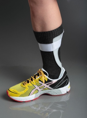
There are methods to improve the foot drop such as: use of splinting in a solid ankle-foot orthoses or foot-up splint. These work to increase the amount of dorsiflexion the foot is held in during gait and can prevent falls as the toes do not get caught on the floor.
Graded exercises to encourage active dorsiflexion has been shown to prevent atrophy and speed up recovery but more research is needed.[2]
Electro-stimulation of the effected muscle groups has also been shown to improve recovery times.[2]
In extreme cases tibialis posterior can be transposed to regain active dorsiflexion through surgery.[3]
Differential Diagnosis[edit | edit source]
add text here relating to the differential diagnosis of this condition
Resources[edit | edit source]
add appropriate resources here
References[edit | edit source]
- ↑ Palastanga N & Soames R Anatomy and Human Movement, Structure and Function. 6th ed. China: Elsevier(Churchill Livingstone) Limited; 2012.
- ↑ 2.0 2.1 2.2 2.3 2.4 Park JH, Restrepo C, Norton R, Mandel S, Sharkey PF, Parvizi J. Common peroneal nerve palsy following total knee arthroplasty: prognostic factors and course of recovery. The Journal of arthroplasty. 2013 Oct 1;28(9):1538-42
- ↑ 3.0 3.1 Baima J, Krivickas L. Evaluation and treatment of peroneal neuropathy. Current reviews in musculoskeletal medicine. 2008 Jun 1;1(2):147-53.
- ↑ Henrichs A. A review of knee dislocations. Journal of athletic training. 2004 Oct;39(4):365.
- ↑ NEJM video. Right foot drop in amublating patient. Available from: https://www.youtube.com/watch?v=cwZYuVB595Q [last accessed 25/08/2013]


