First Rib: Difference between revisions
(Content) |
(Content) |
||
| Line 5: | Line 5: | ||
</div> | </div> | ||
== Description == | == Description == | ||
The first rib is the most superior of the twelve ribs. It is an atypical rib and is an important anatomical landmark. It is one of the borders of the superior thoracic aperture.<ref name=":0">Ramsaroop L, Partab P, Singh B, Satyapal KS. Thoracic origin of a sympathetic supply to the upper limb: the 'nerve of Kuntz' revisited. J Anat. 2001;199:675Y682</ref> | |||
The ribs form the main structure of the thoracic cage that protects the thoracic organs. There are 12 pairs of ribs which are separated by intercostal spaces. The first seven ribs progressively increase in length, the lower five ribs then begin to decrease in length. Ribs are highly vascular and trabecular with a thin outer layer of compact bone. Similar to the first rib, the 11th and 12th ribs are considered atypical ribs due to their anatomical features<ref>https://radiopaedia.org/articles/ribs</ref>. The remaining ribs are typical. | The ribs form the main structure of the thoracic cage that protects the thoracic organs. There are 12 pairs of ribs which are separated by intercostal spaces. The first seven ribs progressively increase in length, the lower five ribs then begin to decrease in length. Ribs are highly vascular and trabecular with a thin outer layer of compact bone. Similar to the first rib, the 11th and 12th ribs are considered atypical ribs due to their anatomical features<ref>https://radiopaedia.org/articles/ribs</ref>. The remaining ribs are typical. | ||
| Line 12: | Line 12: | ||
== Anatomy == | == Anatomy == | ||
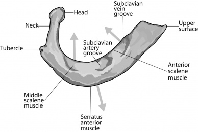
When compared to a typical rib, the first rib is short and thick and only has a single articular facet for the costovertebral joint. The first rib has a head, neck and shaft but lacks a discrete angle<ref>Marhold F, Izay B, Zacherl J, Tschabitscher M, Neumayer C. Thoracoscopic and anatomic landmarks of Kuntz's nerve: implications for sympathetic surgery. Ann Thorac Surg. 2008;86:1653Y1658.</ref>. The shaft is indented laterally, the groove for the subclavian artery, which contains the lowest brachial plexus trunk as well as the subclavian artery. Anterior to the scalene tubercle is another groove for the subclavian vein. There is no costal groove on its inferior surface. It has two tubercles: | When compared to a typical rib, the first rib is short and thick and only has a single articular facet for the costovertebral joint. The first rib has a head, neck and shaft but lacks a discrete angle<ref>Marhold F, Izay B, Zacherl J, Tschabitscher M, Neumayer C. Thoracoscopic and anatomic landmarks of Kuntz's nerve: implications for sympathetic surgery. Ann Thorac Surg. 2008;86:1653Y1658.</ref>. The shaft is indented laterally, the groove for the subclavian artery, which contains the lowest brachial plexus trunk as well as the subclavian artery. Anterior to the scalene tubercle is another groove for the subclavian vein. There is no costal groove on its inferior surface. It has two tubercles: | ||
* | * transverse tubercle: posterior and lateral to the neck; bears an articular facet for the transverse process of T1 | ||
* | * scalene tubercle: anteriorly between the grooves for the subclavian artery and vein; anterior scalene muscle inserts here | ||
** it is also known as | ** it is also known as the Lisfranc tubercle, described by Lisfranc in 1815 | ||
== Blood Supply == | == Blood Supply == | ||
Revision as of 16:25, 21 January 2018
Original Editor -
Top Contributors - Kim Jackson, Maram Salem, Adam Vallely Farrell, Lucinda hampton, Kirenga Bamurange Liliane, Evan Thomas, Mahbubur Rahman, Kai A. Sigel and WikiSysop
Description[edit | edit source]
The first rib is the most superior of the twelve ribs. It is an atypical rib and is an important anatomical landmark. It is one of the borders of the superior thoracic aperture.[1]
The ribs form the main structure of the thoracic cage that protects the thoracic organs. There are 12 pairs of ribs which are separated by intercostal spaces. The first seven ribs progressively increase in length, the lower five ribs then begin to decrease in length. Ribs are highly vascular and trabecular with a thin outer layer of compact bone. Similar to the first rib, the 11th and 12th ribs are considered atypical ribs due to their anatomical features[2]. The remaining ribs are typical.
Anatomy[edit | edit source]
When compared to a typical rib, the first rib is short and thick and only has a single articular facet for the costovertebral joint. The first rib has a head, neck and shaft but lacks a discrete angle[3]. The shaft is indented laterally, the groove for the subclavian artery, which contains the lowest brachial plexus trunk as well as the subclavian artery. Anterior to the scalene tubercle is another groove for the subclavian vein. There is no costal groove on its inferior surface. It has two tubercles:
- transverse tubercle: posterior and lateral to the neck; bears an articular facet for the transverse process of T1
- scalene tubercle: anteriorly between the grooves for the subclavian artery and vein; anterior scalene muscle inserts here
- it is also known as the Lisfranc tubercle, described by Lisfranc in 1815
Blood Supply[edit | edit source]
Arterial blood supply arises from the internal thoracic and superior intercostal arteries. The internal thoracic artery supplies the anterior body wall and its associated structures from the clavicles to the umbilicus. It originates from the first part of the subclavian artery in the base of the neck. The superior intercostal arteries are formed as a direct result of the embryological development of the intersegmental arteries. These arteries are paired structures of the upper thorax which normally form to provide blood flow to the first and second intercostal arteries.[4]
Venous drainage is to the intercostal veins.
Innervation[edit | edit source]
The first rib is innervated by the first intercostal nerve. The intercostal nerves are part of the somatic nervous system, and arise from the anterior rami of the thoracic spinal nerves from T1 to T11. The intercostal nerves are distributed chiefly to the thoracic pleura and abdominal peritoneum and differ from the anterior rami of the other spinal nerves in that each pursues an independent course without plexus formation.[1] The first intercostal nerve is joined to the brachial plexus through a branch, which is equivalent to the lateral cutaneous branches of remaining intercostal nerves. Another exception with the first intercostal nerve is that there is no anterior cutaneous branch. It is also very small as compared to the remaining nerves[5].
Attachments[edit | edit source]
The first rib has several attachments which are listed below;
- anterior scalene muscle: scalene tubercle
- middle scalene muscle: between groove for the subclavian artery and transverse tubercle
- intercostal muscles: from the outer border
- subclavius muscle: arises from the distal shaft and first costal cartilage
- first digitation of the serratus anterior muscle
- parietal pleura: from the inner border
- costoclavicular ligament: anterior to the groove for the subclavian vein
Palpation[edit | edit source]
The first rib is often noted as the most difficult rib to palpate. To palpate the first rib, find the superior border of the upper trapezius muscle and then drop off it anteriorly and direct your palpatory pressure inferiorly against the first rib. Asking a patient to take in a deep breath will elevate the first rib up against your palpating fingers and make palpation easier
Examination[edit | edit source]
First Rib Assessment on hypomobility in Supine:
First Rib Assessment on hypomobility in Prone:
First Rib Assessment on hypomobility in Sitting:
Pathology/Injury[edit | edit source]
References[edit | edit source]
- ↑ 1.0 1.1 Ramsaroop L, Partab P, Singh B, Satyapal KS. Thoracic origin of a sympathetic supply to the upper limb: the 'nerve of Kuntz' revisited. J Anat. 2001;199:675Y682
- ↑ https://radiopaedia.org/articles/ribs
- ↑ Marhold F, Izay B, Zacherl J, Tschabitscher M, Neumayer C. Thoracoscopic and anatomic landmarks of Kuntz's nerve: implications for sympathetic surgery. Ann Thorac Surg. 2008;86:1653Y1658.
- ↑ https://radiopaedia.org/articles/supreme-intercostal-arteries
- ↑ http://www.mananatomy.com/body-systems/nervous-system/intercostal-nerves
- ↑ Physiotutors. First Rib Assessment in Supine | Rib Hypomobility Available from: https://www.youtube.com/watch?v=dPexFNjZB0Q
- ↑ Physiotutors. First Rib Assessment in Prone | Rib Hypomobility. Available from: https://www.youtube.com/watch?v=gfIy_SZ1KMc
- ↑ Physiotutors. First Rib Assessment in Sitting | Rib Hypomobility. Available from: https://www.youtube.com/watch?v=TTMQh4dZqeg


