First Ray: Difference between revisions
Matthew Chin (talk | contribs) No edit summary |
Matthew Chin (talk | contribs) No edit summary |
||
| Line 5: | Line 5: | ||
</div> | </div> | ||
== Introduction == | == Introduction == | ||
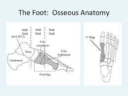
The first ray is the segment of the foot composed of the first metatarsal and first cuneiform bones.<ref name=":0">Glasoe W, Yack J, Saltzman C. Anatomy and Biomechanics of the First Ray[[https://academic.oup.com/ptj/article/79/9/854/2837102]]. Physical Therapy. 1999;79:854-859</ref> The location of this joint is important as it intersects the transverse and medial longitudinal arches.<ref name=":1">D'Amico JC. Understanding the First Ray[[https://www.podiatrym.com/cme/CME1_916.pdf]]. Podiatry Management 2016:109-122.</ref> This segment serves as a critical element in the structural integrity of the foot.<ref name=":0" /> | The '''first ray''' is the segment of the foot composed of the first metatarsal and first cuneiform bones.<ref name=":0">Glasoe W, Yack J, Saltzman C. Anatomy and Biomechanics of the First Ray[[https://academic.oup.com/ptj/article/79/9/854/2837102]]. Physical Therapy. 1999;79:854-859</ref> The location of this joint is important as it intersects the transverse and medial longitudinal arches.<ref name=":1">D'Amico JC. Understanding the First Ray[[https://www.podiatrym.com/cme/CME1_916.pdf]]. Podiatry Management 2016:109-122.</ref> This segment serves as a critical element in the structural integrity of the foot.<ref name=":0" /> | ||
== Anatomy == | == Anatomy == | ||
| Line 22: | Line 22: | ||
=== Hypermobile First Ray === | === Hypermobile First Ray === | ||
Hypermobility at the first ray causes a collapse of the structural framework of the medial longitudinal arch, decreasing the ability of the foot to become a rigid lever required for propulsion. Excessive mobility in the first ray has been implicated in foot pathologies such as hallux valgus, metatarsus varus, and [https://www.physio-pedia.com/Plantar_Fasciitis plantar fasciitis].<ref name=":1" /> | Hypermobility at the first ray causes a collapse of the structural framework of the medial longitudinal arch, decreasing the ability of the foot to become a rigid lever required for propulsion. Excessive mobility in the first ray has been implicated in foot pathologies such as [https://physio-pedia.com/Hallux_Valgus?utm_source=physiopedia&utm_medium=search&utm_campaign=ongoing_internal hallux valgus], metatarsus varus, and [https://www.physio-pedia.com/Plantar_Fasciitis plantar fasciitis].<ref name=":1" /> | ||
== References == | == References == | ||
Revision as of 15:14, 22 September 2020
Original Editor - Matthew Chin
Top Contributors - Matthew Chin and Abbey Wright
Introduction[edit | edit source]
The first ray is the segment of the foot composed of the first metatarsal and first cuneiform bones.[1] The location of this joint is important as it intersects the transverse and medial longitudinal arches.[2] This segment serves as a critical element in the structural integrity of the foot.[1]
Anatomy[edit | edit source]
The first metatarsal is the shortest, strongest, and most important weight-bearing point in the forefoot.[2] In standing, this bone carries 40% of body weight.[2]
The stability of the first metatarsal-cuneiform joint is supported by the tendinous attachments onto the first ray from the tibialis posterior, tibialis anterior, and peroneus longus muscles.[2] Additionally, the two sesamoid bones encased in the tendons beneath the head of the first metatarsal function to elevate the first ray.[1]
Function[edit | edit source]
The first ray serves numerous purposes, including: resisting ground reaction forces, maintaining medial longitudinal arch integrity during midstance supination, allowing first metatarsal head to plantarflex at heel lift, and providing medial stability for propulsive phase.[2]
Pathomechanics[edit | edit source]
Hypomobile First Ray[edit | edit source]
Hypomobility into dorsiflexion of the first ray may cause higher plantar pressure beneath the first ray, which may increase the risk of ulceration of the fat pad under the first metatarsal head in individuals with insensitivity associated with diabetes.[1]
Hypermobile First Ray[edit | edit source]
Hypermobility at the first ray causes a collapse of the structural framework of the medial longitudinal arch, decreasing the ability of the foot to become a rigid lever required for propulsion. Excessive mobility in the first ray has been implicated in foot pathologies such as hallux valgus, metatarsus varus, and plantar fasciitis.[2]


