First Ray: Difference between revisions
Matthew Chin (talk | contribs) No edit summary |
Matthew Chin (talk | contribs) No edit summary |
||
| Line 8: | Line 8: | ||
== Anatomy == | == Anatomy == | ||
The relevant soft tissues surrounding the first ray include the plantar fascia, ligaments, with tendinous attachments onto the first ray of the tibialis posterior, tibialis anterior, and peroneus longus muscles. | |||
[[File:First ray.jpeg]] | [[File:First ray.jpeg]] | ||
Revision as of 21:04, 16 September 2020
Original Editor - User Name
Top Contributors - Matthew Chin and Abbey Wright
Introduction[edit | edit source]
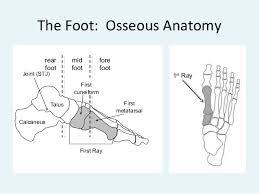
The first ray is the segment of the foot composed of the first metatarsal and first cuneiform bones.[1] The location of this joint is important as it intersects the transverse and medial longitudinal arches.[2] This segment serves as a critical element in the structural integrity of the foot.[1]
Anatomy[edit | edit source]
The relevant soft tissues surrounding the first ray include the plantar fascia, ligaments, with tendinous attachments onto the first ray of the tibialis posterior, tibialis anterior, and peroneus longus muscles.
Biomechanics[edit | edit source]
The first ray serves numerous purposes, including: resisting ground reaction forces, maintaining medial longitudinal arch integrity during midstance supination, allowing first metatarsal head to plantarflex at heel lift, and providing medial stability for propulsive phase.[2]


