File:Diverticula.jpg: Difference between revisions
No edit summary |
No edit summary |
||
| Line 3: | Line 3: | ||
Haymanj - Self-photographed | Haymanj - Self-photographed | ||
== Licensing == | == Licensing == | ||
{{Template:Radiopedia}} | {{Template:Radiopedia}} | ||
Revision as of 06:02, 19 March 2024
Summary[edit | edit source]
Large bowel (sigmoid colon) showing multiple diverticula. Note how the diverticula appear on either side of the longitudinal muscle bundle (taenium).
Haymanj - Self-photographed
Licensing[edit | edit source]
Please include the following information in the summary box:
Image courtesy of Dr [name of contributor], Radiopaedia.org; rID:xxxx (which can be found top right in the "case information" box on case pages).
Radiopaedia.org, the copyright holder, gave special permission to use this work in Physiopedia. This permission does not extend to any other person or organisation. Please do not use this image without permission from the copyright holder.
File history
Click on a date/time to view the file as it appeared at that time.
| Date/Time | Thumbnail | Dimensions | User | Comment | |
|---|---|---|---|---|---|
| current | 03:32, 6 April 2016 | 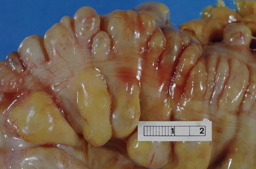 | 1,024 × 678 (100 KB) | Katie Countryman (talk | contribs) | Large bowel (sigmoid colon) showing multiple diverticula. Note how the diverticula appear on either side of the longitudinal muscle bundle (taenium). Haymanj - Self-photographed |
You cannot overwrite this file.
File usage
The following page uses this file:

