Eye Muscle Exercise
This article or area is currently under construction and may only be partially complete. Please come back soon to see the finished work! (Template:28/Template:5/Template:2020)
Introduction[edit | edit source]
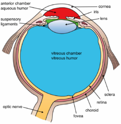
The eye divided into three layers; the outermost layer is a fibrous layer and it is consists of the cornea that is transparent located at the center of the eye, the sclera is white and covers the rest of the eye. The second layer is a vascular layer it is consists of the choroid (contain blood supply to the retina), the iris (contains pupils and smooth muscles that controls its diameter[1]), and the ciliary body which is a tissue extends from the sclera and attaches to the lens via suspensory ligament. The inner layer and it contains the retina.
The extraocular muscles are seven muscles supplied by cranial nerves that are responsible for eye movement, in some cases when these nerves are affected consequently they affect eye movements.
Eye Muscles[edit | edit source]
The ocular movement occurs around the three axis[2]:
- Adduction (medial pupil movement toward nose)/ Abduction (lateral pupil movement) around the vertical axis.
- Elevation (superior movement)/ Depression (inferior movement) around the transverse axis.
- Intorsion/ Extorsion (the movement away and toward nose) that we need during tilting the head around the anteroposterior axis
The seven muscles of extraocular muscles divided into 4 recti, 2 obliques muscles, and one levator palpebrae superiors that are responsible for elevation of the superior eyelid.
| Muscle | Origin | Insertion | Nerve supply | Action |
|---|---|---|---|---|
| Superior rectus | common tendinous ring | superior and anterior aspect of sclera | oculomotor nerve (cranial nerve III) | elevation and contributes to adduction and intorsion |
| Inferior rectus | inferior and anterior aspect of sclera | depression and contributes to adduction and extorsion | ||
| Medial rectus | medial aspect of sclera | adducts eye | ||
| Lateral rectus | lateral aspect of sclera | abducens nerve (cranial nerve VI) | abducts eye | |
| Superior oblique | body of sphenoid bone | at sclera posterior to superior rectus | Trochlear nerve cranial nerve IV) | abduction, depression, and intorsion of eye |
| Inferior oblique | anterior aspect of orbital floor | at sclera posterior to lateral rectus. | Oculomotor nerve (cranial nerve III) | abduction, elevation, and extortion of eye |
| Levator palpebrae superiors | sphenoid bone | superior eyelid | oculomotor nerve (cranial nerve III) | elevation of the superior eyelid. |
Clinical significance[edit | edit source]
Extraocular muscle paralysis may happen due to injury or disease according to the cranial nerve that will be affected, palsy of the oculomotor nerve will affect the majority of extraocular muscles and the eye ill adopted in a down and out position. Palsy of the abducens nerve will affect the lateral rectus and the eye will be addicted by medial rectus. If the trochlear nerve is affected the patient will complain of diplopia[3].
Strabismus, due to abnormalities in neuromuscular control weakness or injury to the inferior rectus muscle may be involved.[4]
Assessment of extraocular muscles[edit | edit source]
Assessment of extraocular muscle function can be done by asking the patient to look in nine directions by following the clinician's finger as he draws ''H'' in the air. The patient will look at up, down, right, left, up and right, up and left, down and right, down and left.[4]
Ocular alignment is tested by several methods, for example, corneal-light reflex.
Eye muscles exercises[edit | edit source]
Who benefit from exercises:
Digital eye strain as in people who work on a computer for along time and that may cause dry eyes, eye strain, blurred vision, and headaches.
Increased light sensitivity.
A surgery that need strength the muscle
If there is a problem in focusing eyes to read.
Lazy eye that develops in some children, eye exercises stimulate the vision centers in the brain.
Patients with astigmatism, myopia, dyslexia, paralyzed eye muscle won't benefit from eye exercises[5][6].
Resources[edit | edit source]
References[edit | edit source]
- ↑ Rehman I, Hazhirkarzar B, Patel BC. Anatomy, head and neck, eye.
- ↑ Rehman I, Hazhirkarzar B, Patel BC. Anatomy, head and neck, eye.
- ↑ https://teachmeanatomy.info/head/organs/eye/extraocular-muscles/
- ↑ 4.0 4.1 Shumway CL, Motlagh M, Wade M. Anatomy, head and neck, eye extraocular muscles. InStatPearls [Internet] 2019 Jun 26. StatPearls Publishing.
- ↑ https://www.healthline.com/health/eye-health/eye-exercises
- ↑ https://www.webmd.com/eye-health/eye-exercises


