Chondrosarcoma
Original Editors - Students from Bellarmine University's Pathophysiology of Complex Patient Problems project.
Top Contributors - Dalton O'Brien, Corey Hardesty, Lucinda hampton, Nikhil Benhur Abburi, 127.0.0.1, Admin, Elaine Lonnemann, WikiSysop, Kim Jackson, Vidya Acharya and Rewan Elsayed Elkanafany
Definition/Description[edit | edit source]
Chondrosarcoma is a type of malignant bone tumor that most commonly effects the pelvis, sternum, scapula, or cartilage of long bones in the extremities. Typically, chondrosarcoma forms within bone or cartilage cells around the articular surfaces and can be either slow-growing and form spontaneously, or due to malignant changes in a preexisting (secondary) bone tumorCite error: Invalid <ref> tag; name cannot be a simple integer. Use a descriptive title.
There are three variants of chondrosarcoma which have different prognosis for each, include:Cite error: Invalid <ref> tag; name cannot be a simple integer. Use a descriptive title
- Dedifferentiated chondrosarcoma- occurs in older patients and is considered a more aggressive tumor
- Clear cell chondrosarcoma- rare, slow-growing and typically do not metastasize
- Mesenchymal chondrosarcoma- grow rapidly and are more responsive to chemotherapy and radiation treatments
Prevalence[edit | edit source]
Chondrosarcoma is the most common malignant cartilage tumor and the third most common bone sarcoma (behind osteosarcoma and Ewing's sarcoma). This pathology commonly occurs in adults between the ages of 20-70, males slightly more affected than females, with peak occurrences in the fifth and seventh decades. It is possible to develop chondrosarcoma at younger age groups, which usually leads to higher malignancy and metastases rates. The sites most often affected are the proximal femur, followed by the proximal humerus.Cite error: Invalid <ref> tag; name cannot be a simple integer. Use a descriptive titleCite error: Invalid <ref> tag; name cannot be a simple integer. Use a descriptive title
Characteristics/Clinical Presentation[edit | edit source]
Clinical features seen in patients with chondrosasrcoma include age >25 years old, inflammatory pain (pain with palpation), axial skelton involvement.Cite error: Invalid <ref> tag; name cannot be a simple integer. Use a descriptive titleCite error: Invalid <ref> tag; name cannot be a simple integer. Use a descriptive title
Other characteristics of chondrosarcomaCite error: Invalid <ref> tag; name cannot be a simple integer. Use a descriptive title:
- Palpable mass
- Back, pelvis, or thigh pain
- Sciatica
- Bladder symptoms
- Unilateral edema
Associated Co-morbidities
[edit | edit source]
Sarcomas that involve soft tissue, such as chondrosarcoma, are more commonly found in individuals who have:Cite error: Invalid <ref> tag; name cannot be a simple integer. Use a descriptive title
- von Recklinghausen's disease
- Gardner's syndrome
- Werner's syndrome
- Tuberous sclerosis
- Basal cell nevus syndrome
- Li-Fraumeni syndrome
- AIDS (Kaposi's sarcoma)
- Radiation (following surgery), herbicides (phenoxyacetic acid), wood preservatives (chlorophenols), or vinyl chloride exposure
Medications[edit | edit source]
Targeted therapy for bone cancers include Imatinib and Denosumab, however there are no medications directly related to treating chondrosarcoma specifically, beyond traditional chemotherapyCite error: Invalid <ref> tag; name cannot be a simple integer. Use a descriptive titleCite error: Invalid <ref> tag; name cannot be a simple integer. Use a descriptive title. Following surgery, Hydrocodone can be prescribed for pain to the patientsCite error: Invalid <ref> tag; name cannot be a simple integer. Use a descriptive title.
Diagnostic Tests/Lab Tests/Lab Values[edit | edit source]
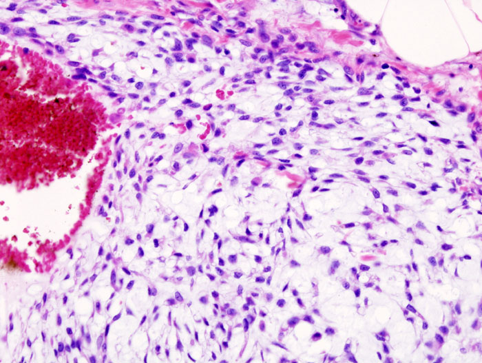
Chondrosarcoma is initially assessed for aggressiveness via radiographs. CT and MRI are used to view the amount of cortical loss seen. A biopsy is later performed to confirm the diagnosis. Pathologists follow up with histology to grade the tumor.
Histological Grades:
- Grade 1: low grade neoplasm; more benign; no cortical expansion, destruction, or soft tissue mass on imaging (90% of patients exhibit a 5-year survival rate)
- Grades 2-3: high grade neoplasm; imaging reveals cortical expansion, destruction, and/or soft tissue mass (40-60% of patients exhibit a 5-year survival rate)
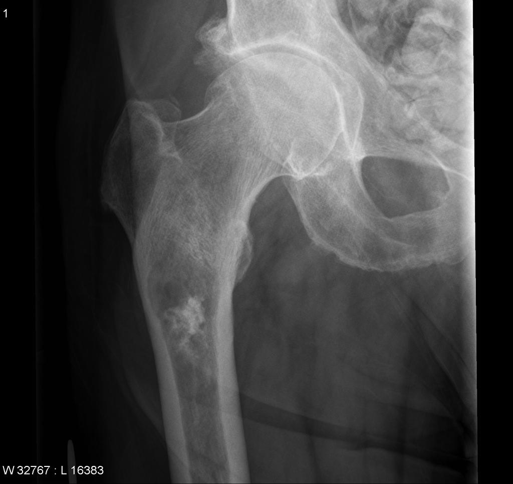
Radiological features of Chondrosarcoma include:
- Intramedullary lesion
- Lesion >5cm
- Microfractures
- Endosteal scalloping (focal resorption of inner margin of cortical bone)
- Loss of calcification
- Soft tissue mass
Cite error: Invalid <ref> tag; name cannot be a simple integer. Use a descriptive titleCite error: Invalid <ref> tag; name cannot be a simple integer. Use a descriptive title
Intramedullary lesion with calcification.Cite error: Invalid <ref> tag; name cannot be a simple integer. Use a descriptive title
Histopathogic image of chondrosarcomaCite error: Invalid <ref> tag; name cannot be a simple integer. Use a descriptive title
Causes[edit | edit source]
As a whole, the epidemiology of soft tissue sarcomas is not well understood. Although there is no proven genetic disposition, some studies indicate exposure to certain herbicides, wood preservatives, and radiation lead to an increased risk of development. Currently, chrondrosarcomas are thought to form spontaneously or from preexisting bone tumors that have become malignant. Benign bone tumors (osteochondromas, enchondromas, chondromas) that become malignant are referred to as secondary chondrosarcomas and are still composed of cartilaginous tissue entirelyCite error: Invalid <ref> tag; name cannot be a simple integer. Use a descriptive title.
Systemic Involvement[edit | edit source]
Haversian system and bone marrow invasion
Periosteal reaction
Occasional sites of necrosis and hemorrhage
Can affect pulmonary system via metastasis to lungs, trachea, or chest wallCite error: Invalid <ref> tag; name cannot be a simple integer. Use a descriptive titleCite error: Invalid <ref> tag; name cannot be a simple integer. Use a descriptive title
Medical Management (current best evidence)[edit | edit source]
Following a biopsy to confirm the diagnosis, surgery is the most common procedure to remove the tumor. Even for higher grade tumors, limb-sparing curettage (via cryotherapy) is more common, with amputation occurring in rare instances.
Unlike most cancers, typical chondrosarcoma does not respond to chemotherapy and is resistant to radiation therapy. Different forms of chondrosarcoma including dedifferentiated and mesenchymal may be trial chemotherapy before or after surgery and follow a similar protocol to osteosarcoma and Ewing's sarcoma respectively. Depending on the grading of the sarcoma, proton-beam radiation has had some success, but does require very high doses and is considered less frequently than surgical approaches.Cite error: Invalid <ref> tag; name cannot be a simple integer. Use a descriptive titleCite error: Invalid <ref> tag; name cannot be a simple integer. Use a descriptive title
Physical Therapy Management (current best evidence)[edit | edit source]
Physical therapy management will most often occur following surgery to excise the tumor. Depending on the site of the lesion, the surgeon may have different protocols, but physical therapy can focus on treatments that will decrease pain, decrease edema, and improve patients' quality of life.
Following surgery, an acute care physical therapist will teach the patient skills like bed mobility, weight bearing precautions, ambulation, and stair negotitation. In outpatient physical therapy, the patient will undergo manual therapy and soft tissue mobilization to improve tissue extensibility, reduce edema, and improve range of motion. The patient will also perform therapuetic exercises to increase range of motion and muscle strength to address deficits normally seen following surgery. Gait training will continue to be incorportated and adapted to the changing weight bearing precautions set forth by the surgeon. As the patient progresses, the treatments will progress to become more functional and incorporate activites that are important to the patient.Cite error: Invalid <ref> tag; name cannot be a simple integer. Use a descriptive title
Alternative/Holistic Management (current best evidence)[edit | edit source]
Currently, there are no evidence based, documented reports of alternative/holistic management for chondrosarcoma.
Differential Diagnosis[edit | edit source]
Chondrosarcoma presents similarly to other musculoskeletal injuries/illnesses of the affected joint. For example, a patient with chondrosarcoma of the hip will complain of intermittenet anterior thigh/hip pain. The pain can present as either hip joint in origin or nonarticular hip origin. Other diagnoses with pain of hip joint origin include osseus necrosis, stress fracture, hip dysplasia, or intra-articular pathology (labral tear, ligamentum teres tear, or loose body in the joint). Diagnoses of nonarticular hip origin of pain include bursitis, tendonopathy, muscle strain, urogenital conditions, metabolic disease, vascular conditions, or infection.Cite error: Invalid <ref> tag; name cannot be a simple integer. Use a descriptive title
Case Reports[edit | edit source]
Ferrer-Santacreu E, Ortiz-Cruz E, Díaz-Almirón M, Pozo Kreilinger J. Enchondroma versus Chondrosarcoma in Long Bones of Appendicular Skeleton: Clinical and Radiological Criteria—A Follow-Up. Journal Of Oncology [serial on the Internet]. (2016, Feb 23), [cited April 10, 2016]; 1-10. Available from: Academic Search Complete.
Resources
[edit | edit source]
What is bone cancer? [Internet]. Cancer.org. 2016 [cited 10 April 2016]. Available from: http://www.cancer.org/cancer/bonecancer/detailedguide/bone-cancer-what-is-bone-cancer
Treating specific bone cancers [Internet]. Cancer.org. 2016 [cited 10 April 2016]. Available from: http://www.cancer.org/cancer/bonecancer/detailedguide/bone-cancer-treating-treating-specific-bone-cancers
National Cancer Institute [Internet]. Cancer.gov. 2016 [cite 10 April 2016]. Available from: http://www.cancer.gov/
Recent Related Research (from Pubmed)[edit | edit source]
Failed to load RSS feed from http://www.ncbi.nlm.nih.gov/entrez/eutils/erss.cgi?rss_guid=1fuD0jDVLZMdoQ3wW8sTZdXu_YpsmeiAkYlYZ33CDYyy4P89sG|charset=UTF-8|short|max=10: Error parsing XML for RSS
References[edit | edit source]
see adding references tutorial.


