Cerebellum: Difference between revisions
(text) |
(added video and text) |
||
| Line 9: | Line 9: | ||
</div> | </div> | ||
== Introduction == | == Introduction == | ||
The cerebellum is a vital component in the human brain as it plays a role in motor movement regulation and balance control. The cerebellum <ref>Jimsheleishvili S, Dididze M. [https://www.ncbi.nlm.nih.gov/books/NBK538167/ Neuroanatomy, Cerebellum.] InStatPearls [Internet] 2019 Mar 2. StatPearls Publishing.Available from:https://www.ncbi.nlm.nih.gov/books/NBK538167/ (last accessed 19.1.2020)</ref> | [[File:Cerebellum gif.gif|right|frameless|400x400px]] | ||
The cerebellum is a vital component in the human brain as it plays a role in motor movement regulation and balance control. The cerebellum <ref name=":1">Jimsheleishvili S, Dididze M. [https://www.ncbi.nlm.nih.gov/books/NBK538167/ Neuroanatomy, Cerebellum.] InStatPearls [Internet] 2019 Mar 2. StatPearls Publishing.Available from:https://www.ncbi.nlm.nih.gov/books/NBK538167/ (last accessed 19.1.2020)</ref> (see image on R, horizontal fissure marked red) | |||
* coordinates gait | * coordinates gait | ||
* maintains posture, | * maintains posture, | ||
| Line 32: | Line 32: | ||
There are three anatomical lobes that can be distinguished in the cerebellum; the anterior lobe, the posterior lobe and the flocculonodular lobe. These lobes are divided by two fissures – the primary fissure and posterolateral fissure. | There are three anatomical lobes that can be distinguished in the cerebellum; the anterior lobe, the posterior lobe and the flocculonodular lobe. These lobes are divided by two fissures – the primary fissure and posterolateral fissure. | ||
[[File:Cerebellum1.png|right|frameless|380x380px]] | |||
2. Zones | 2. Zones | ||
| Line 45: | Line 45: | ||
* Vestibulocerebellum – the functional equivalent to the flocculonodular lobe. It is involved in controlling balance and ocular reflexes, mainly fixation on a target. It receives inputs from the vestibular system, and sends outputs back to the vestibular nuclei. | * Vestibulocerebellum – the functional equivalent to the flocculonodular lobe. It is involved in controlling balance and ocular reflexes, mainly fixation on a target. It receives inputs from the vestibular system, and sends outputs back to the vestibular nuclei. | ||
== Function == | == Function == | ||
The | Function by regions | ||
* The cortex of the vermis coordinates the movements of the trunk, including the neck, shoulders, thorax, abdomen, and hips. | |||
* Control of the distal extremity muscles is by the intermediate zone of cerebellar hemispheres, located adjacent to the vermis. | |||
* The remaining lateral area of each cerebellar hemisphere provides the planning of sequential movements of the entire body along with involvement in the conscious assessment of movement errors | |||
Main Functions overall<ref name=":2">Lumenlearning. [[Cerebellum]] Available from:https://courses.lumenlearning.com/boundless-ap/chapter/the-cerebellum/ (last accessed 20.1.2020)</ref> | |||
[[File:Cerebellum role as a comparator.png|right|frameless|450x450px]] | |||
* The cerebellum is essential for making fine adjustments to motor actions. Cerebellar dysfunction primarily results in problems with motor control. | * The cerebellum is essential for making fine adjustments to motor actions. Cerebellar dysfunction primarily results in problems with motor control. | ||
* Four principles are important to cerebellar processing: feedforward processing, divergence and convergence, modularity, and plasticity. | * Four principles are important to cerebellar processing: feedforward processing, divergence and convergence, modularity, and plasticity. | ||
| Line 55: | Line 58: | ||
* The cerebellar system is divided into thousands of independent modules with similar structure. | * The cerebellar system is divided into thousands of independent modules with similar structure. | ||
== Blood Supply == | |||
[[File:Circle of Willis en.svg.png|right|frameless]] | |||
The cerebellum receives its blood supply from three paired arteries (originate from the vertebrobasilar anterior system)<ref name=":0" /> | |||
* Superior cerebellar artery (SCA) | |||
* Anterior inferior cerebellar artery (AICA) | |||
* Posterior inferior cerebellar artery (PICA) | |||
The SCA and AICA are branches of the basilar artery, which wraps around the anterior aspect of the pons before reaching the cerebellum. The PICA is a branch of the vertebral artery. | |||
Venous drainage of the cerebellum is by the superior and inferior cerebellar veins. They drain into the superior petrosal, transverse and straight dural venous sinuses. | |||
== Clinical Significance == | |||
The cerebellum receives afferent information about voluntary muscle movements from the cerebral cortex and from the muscles, tendons, and joints. It also receives information concerning balance from the vestibular nuclei. Each cerebellar hemisphere controls the same side of the body, thus if damaged the symptoms will occur ipsilaterally.<ref name=":1" /> | |||
Dysfunction of the cerebellum can produce a wide range of symptoms and signs. | |||
The most common cause of cerebellar dysfunction is alcohol poisoning, but also trauma, multiple sclerosis, tumors, thrombosis of the cerebellar arteries and stroke.<ref name=":1" /> | |||
The clinical picture depends on the functional area of the cerebellum that is affected<ref name=":2" />. | |||
* Damage to the flocculonodular lobe (vestibulocerebellum): loss of equilibrium causing an altered walking gait | |||
* Lateral zone damage: problems with skilled voluntary and planned movements leading to errors in intended movements (eg., dysdiadochokinesia, the inability to perform rapid alternating movements). | |||
* Damage to the midline portion: disruption of whole-body movements | |||
* Damage to the upper part of the cerebellum: gait impairments and other problems with leg coordination (ie, ataxia). | |||
A wide variety of manifestations are possible (remembered by acronym ‘DANISH‘)<ref name=":0" /> | |||
* Dysdiadochokinesia - the lack of ability to perform rapidly alternating movements. Ask the patient to quickly supinate and pronate both forearms simultaneously. Movements will be slow and incomplete on the side of the cerebellar lesion.<ref name=":1" /> | |||
* Ataxia - voluntary movement disturbance involves tremor with fine movements eg writing or buttoning the clothes. Finger to nose test is performed to examine the coordination of the muscle movements, testing tip of the nose with the index finger test the movements are not properly coordinated, and tremor is observed at the end of the movement, called intention tremor. A similar test can be performed on the lower limbs ask patient to place the heel of one foot against the shin of the opposite leg.<ref name=":1" /> | |||
* Nystagmus (coarse) - Ataxia of ocular muscles, a rhythmical oscillation of the eyes. To provoke nystagmus, the patient should rotate eyes horizontally | |||
* Intention tremor | |||
* Dysarthria/Scanning speech - ataxia of the larynx muscles, speech is slurred and syllables are separated from one another. | |||
* Hypotonia - the muscles lose resistance to palpation due to diminished influence of the cerebellum on gamma motor neurons. The patient walks with a broad-based gait and leans toward the affected side.<ref name=":1" /> | |||
The short video below gives a good picture of these manifestations | |||
{{#ev:youtube|https://www.youtube.com/watch?v=fMMcMPI68V0&app=desktop|width}}<ref>Soton brain hub Cerebellar disease symptoms Available from:https://www.youtube.com/watch?v=fMMcMPI68V0&app=desktop (last accessed 20.1.2020)</ref> | |||
== Resources == | == Resources == | ||
*bulleted list | *bulleted list | ||
Revision as of 07:45, 20 January 2020
This article or area is currently under construction and may only be partially complete. Please come back soon to see the finished work! (20/01/2020)
Original Editor - Your name will be added here if you created the original content for this page.
Top Contributors - Lucinda hampton, Rahma Ahmed Ahmed Bahbah, Rusfid FM, Kim Jackson, Nikhil Benhur Abburi, Khloud Shreif, Mason Trauger and Jonathan Wong
Introduction[edit | edit source]
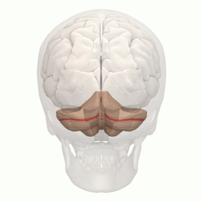
The cerebellum is a vital component in the human brain as it plays a role in motor movement regulation and balance control. The cerebellum [1] (see image on R, horizontal fissure marked red)
- coordinates gait
- maintains posture,
- controls muscle tone and voluntary muscle activity
- is unable to initiate muscle contraction.
Damage to this area in humans results in a loss in the ability to control fine movements, maintain posture, and motor learning
Anatomical Position[edit | edit source]
The cerebellum is located at the back of the brain, immediately inferior to the occipital and temporal lobes, and within the posterior cranial fossa. It is separated from these lobes by the tentorium cerebelli, a tough layer of dura mater.
It lies at the same level of and posterior to the pons, from which it is separated by the fourth ventricle.
Structure[edit | edit source]
The cerebellum consists of two hemispheres which are connected by the vermis, a narrow midline area. The cerebellum consists of grey matter and white matter:[2]
- Grey matter – located on the surface of the cerebellum. It is tightly folded, forming the cerebellar cortex.
- White matter – located underneath the cerebellar cortex. Embedded in the white matter are the four cerebellar nuclei (the dentate, emboliform, globose, and fastigi nuclei).
There are three ways that the cerebellum can be subdivided – anatomical lobes, zones and functional division[2]
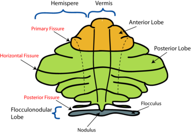
1. Anatomical Lobes
There are three anatomical lobes that can be distinguished in the cerebellum; the anterior lobe, the posterior lobe and the flocculonodular lobe. These lobes are divided by two fissures – the primary fissure and posterolateral fissure.
2. Zones
There are three cerebellar zones. In the midline of the cerebellum is the vermis. Either side of the vermis is the intermediate zone. Lateral to the intermediate zone are the lateral hemispheres. There is no difference in gross structure between the lateral hemispheres and intermediate zones
3. Functional Divisions
The cerebellum can also be divided by function. There are three functional areas of the cerebellum – the cerebrocerebellum, the spinocerebellum and the vestibulocerebellum.
- Cerebrocerebellum – the largest division, formed by the lateral hemispheres. It is involved in planning movements and motor learning. It receives inputs from the cerebral cortex and pontine nuclei, and sends outputs to the thalamus and red nucleus. This area also regulates coordination of muscle activation and is important in visually guided movements.
- Spinocerebellum – comprised of the vermis and intermediate zone of the cerebellar hemispheres. It is involved in regulating body movements by allowing for error correction. It also receives proprioceptive information.
- Vestibulocerebellum – the functional equivalent to the flocculonodular lobe. It is involved in controlling balance and ocular reflexes, mainly fixation on a target. It receives inputs from the vestibular system, and sends outputs back to the vestibular nuclei.
Function[edit | edit source]
Function by regions
- The cortex of the vermis coordinates the movements of the trunk, including the neck, shoulders, thorax, abdomen, and hips.
- Control of the distal extremity muscles is by the intermediate zone of cerebellar hemispheres, located adjacent to the vermis.
- The remaining lateral area of each cerebellar hemisphere provides the planning of sequential movements of the entire body along with involvement in the conscious assessment of movement errors
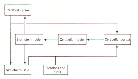
Main Functions overall[3]
- The cerebellum is essential for making fine adjustments to motor actions. Cerebellar dysfunction primarily results in problems with motor control.
- Four principles are important to cerebellar processing: feedforward processing, divergence and convergence, modularity, and plasticity.
- Signal processing in the cerebellum is almost entirely feedforward. Signals move through the system from input to output with very little internal transmission.
- The cerebellum both receives input and transmits output via a limited number of cells.
- The cerebellar system is divided into thousands of independent modules with similar structure.
Blood Supply[edit | edit source]
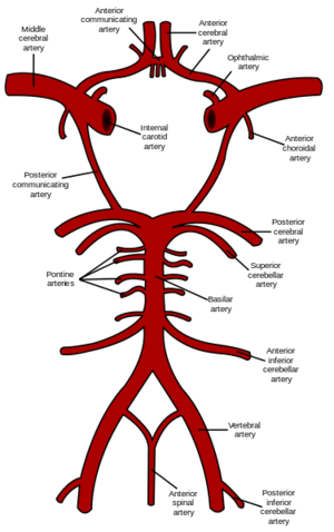
The cerebellum receives its blood supply from three paired arteries (originate from the vertebrobasilar anterior system)[2]
- Superior cerebellar artery (SCA)
- Anterior inferior cerebellar artery (AICA)
- Posterior inferior cerebellar artery (PICA)
The SCA and AICA are branches of the basilar artery, which wraps around the anterior aspect of the pons before reaching the cerebellum. The PICA is a branch of the vertebral artery.
Venous drainage of the cerebellum is by the superior and inferior cerebellar veins. They drain into the superior petrosal, transverse and straight dural venous sinuses.
Clinical Significance[edit | edit source]
The cerebellum receives afferent information about voluntary muscle movements from the cerebral cortex and from the muscles, tendons, and joints. It also receives information concerning balance from the vestibular nuclei. Each cerebellar hemisphere controls the same side of the body, thus if damaged the symptoms will occur ipsilaterally.[1]
Dysfunction of the cerebellum can produce a wide range of symptoms and signs.
The most common cause of cerebellar dysfunction is alcohol poisoning, but also trauma, multiple sclerosis, tumors, thrombosis of the cerebellar arteries and stroke.[1]
The clinical picture depends on the functional area of the cerebellum that is affected[3].
- Damage to the flocculonodular lobe (vestibulocerebellum): loss of equilibrium causing an altered walking gait
- Lateral zone damage: problems with skilled voluntary and planned movements leading to errors in intended movements (eg., dysdiadochokinesia, the inability to perform rapid alternating movements).
- Damage to the midline portion: disruption of whole-body movements
- Damage to the upper part of the cerebellum: gait impairments and other problems with leg coordination (ie, ataxia).
A wide variety of manifestations are possible (remembered by acronym ‘DANISH‘)[2]
- Dysdiadochokinesia - the lack of ability to perform rapidly alternating movements. Ask the patient to quickly supinate and pronate both forearms simultaneously. Movements will be slow and incomplete on the side of the cerebellar lesion.[1]
- Ataxia - voluntary movement disturbance involves tremor with fine movements eg writing or buttoning the clothes. Finger to nose test is performed to examine the coordination of the muscle movements, testing tip of the nose with the index finger test the movements are not properly coordinated, and tremor is observed at the end of the movement, called intention tremor. A similar test can be performed on the lower limbs ask patient to place the heel of one foot against the shin of the opposite leg.[1]
- Nystagmus (coarse) - Ataxia of ocular muscles, a rhythmical oscillation of the eyes. To provoke nystagmus, the patient should rotate eyes horizontally
- Intention tremor
- Dysarthria/Scanning speech - ataxia of the larynx muscles, speech is slurred and syllables are separated from one another.
- Hypotonia - the muscles lose resistance to palpation due to diminished influence of the cerebellum on gamma motor neurons. The patient walks with a broad-based gait and leans toward the affected side.[1]
The short video below gives a good picture of these manifestations
Resources[edit | edit source]
- bulleted list
- x
or
- numbered list
- x
References[edit | edit source]
- ↑ 1.0 1.1 1.2 1.3 1.4 1.5 Jimsheleishvili S, Dididze M. Neuroanatomy, Cerebellum. InStatPearls [Internet] 2019 Mar 2. StatPearls Publishing.Available from:https://www.ncbi.nlm.nih.gov/books/NBK538167/ (last accessed 19.1.2020)
- ↑ 2.0 2.1 2.2 2.3 Teach me anatomy. The Cerebellum Available from: https://teachmeanatomy.info/neuroanatomy/structures/cerebellum/ (last accessed 20.1.2020)
- ↑ 3.0 3.1 Lumenlearning. Cerebellum Available from:https://courses.lumenlearning.com/boundless-ap/chapter/the-cerebellum/ (last accessed 20.1.2020)
- ↑ Soton brain hub Cerebellar disease symptoms Available from:https://www.youtube.com/watch?v=fMMcMPI68V0&app=desktop (last accessed 20.1.2020)


