Cerebellum: Difference between revisions
No edit summary |
(text) |
||
| Line 16: | Line 16: | ||
* is unable to initiate muscle contraction. | * is unable to initiate muscle contraction. | ||
Damage to this area in humans results in a loss in the ability to control fine movements, maintain posture, and motor learning | Damage to this area in humans results in a loss in the ability to control fine movements, maintain posture, and motor learning | ||
== Anatomical Position == | |||
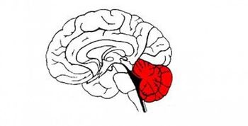
The cerebellum is located at the back of the brain, immediately inferior to the occipital and temporal lobes, and within the posterior cranial fossa. It is separated from these lobes by the tentorium cerebelli, a tough layer of dura mater. | |||
It lies at the same level of and posterior to the pons, from which it is separated by the fourth ventricle. | |||
== Structure == | == Structure == | ||
The cerebellum consists of two hemispheres which are connected by the vermis, a narrow midline area. The cerebellum consists of grey matter and white matter:<ref name=":0">Teach me anatomy. [https://teachmeanatomy.info/neuroanatomy/structures/cerebellum/ The Cerebellum] Available from: https://teachmeanatomy.info/neuroanatomy/structures/cerebellum/ (last accessed 20.1.2020)</ref> | |||
* Grey matter – located on the surface of the cerebellum. It is tightly folded, forming the cerebellar cortex. | |||
* White matter – located underneath the cerebellar cortex. Embedded in the white matter are the four cerebellar nuclei (the dentate, emboliform, globose, and fastigi nuclei). | |||
There are three ways that the cerebellum can be subdivided – anatomical lobes, zones and functional division<ref name=":0" /> | |||
1. Anatomical Lobes | |||
There are three anatomical lobes that can be distinguished in the cerebellum; the anterior lobe, the posterior lobe and the flocculonodular lobe. These lobes are divided by two fissures – the primary fissure and posterolateral fissure. | |||
2. Zones | |||
There are three cerebellar zones. In the midline of the cerebellum is the vermis. Either side of the vermis is the intermediate zone. Lateral to the intermediate zone are the lateral hemispheres. There is no difference in gross structure between the lateral hemispheres and intermediate zones | |||
3. Functional Divisions | |||
The cerebellum can also be divided by function. There are three functional areas of the cerebellum – the cerebrocerebellum, the spinocerebellum and the vestibulocerebellum. | |||
* Cerebrocerebellum – the largest division, formed by the lateral hemispheres. It is involved in planning movements and motor learning. It receives inputs from the cerebral cortex and pontine nuclei, and sends outputs to the thalamus and red nucleus. This area also regulates coordination of muscle activation and is important in visually guided movements. | |||
* Spinocerebellum – comprised of the vermis and intermediate zone of the cerebellar hemispheres. It is involved in regulating body movements by allowing for error correction. It also receives proprioceptive information. | |||
* Vestibulocerebellum – the functional equivalent to the flocculonodular lobe. It is involved in controlling balance and ocular reflexes, mainly fixation on a target. It receives inputs from the vestibular system, and sends outputs back to the vestibular nuclei. | |||
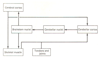
[[File:Cerebellum role as a comparator.png|right|frameless|400x400px]] | [[File:Cerebellum role as a comparator.png|right|frameless|400x400px]] | ||
== Function == | == Function == | ||
The key points being<ref>Lumenlearning. [[Cerebellum]] Available from:https://courses.lumenlearning.com/boundless-ap/chapter/the-cerebellum/ (last accessed 20.1.2020)</ref> | |||
* The cerebellum is essential for making fine adjustments to motor actions. Cerebellar dysfunction primarily results in problems with motor control. | |||
* Four principles are important to cerebellar processing: feedforward processing, divergence and convergence, modularity, and plasticity. | |||
* Signal processing in the cerebellum is almost entirely feedforward. Signals move through the system from input to output with very little internal transmission. | |||
* The cerebellum both receives input and transmits output via a limited number of cells. | |||
* The cerebellar system is divided into thousands of independent modules with similar structure. | |||
== Resources == | == Resources == | ||
Revision as of 07:05, 20 January 2020
This article or area is currently under construction and may only be partially complete. Please come back soon to see the finished work! (20/01/2020)
Original Editor - Your name will be added here if you created the original content for this page.
Top Contributors - Lucinda hampton, Rahma Ahmed Ahmed Bahbah, Rusfid FM, Kim Jackson, Nikhil Benhur Abburi, Khloud Shreif, Mason Trauger and Jonathan Wong
Introduction[edit | edit source]
The cerebellum is a vital component in the human brain as it plays a role in motor movement regulation and balance control. The cerebellum [1]
- coordinates gait
- maintains posture,
- controls muscle tone and voluntary muscle activity
- is unable to initiate muscle contraction.
Damage to this area in humans results in a loss in the ability to control fine movements, maintain posture, and motor learning
Anatomical Position[edit | edit source]
The cerebellum is located at the back of the brain, immediately inferior to the occipital and temporal lobes, and within the posterior cranial fossa. It is separated from these lobes by the tentorium cerebelli, a tough layer of dura mater.
It lies at the same level of and posterior to the pons, from which it is separated by the fourth ventricle.
Structure[edit | edit source]
The cerebellum consists of two hemispheres which are connected by the vermis, a narrow midline area. The cerebellum consists of grey matter and white matter:[2]
- Grey matter – located on the surface of the cerebellum. It is tightly folded, forming the cerebellar cortex.
- White matter – located underneath the cerebellar cortex. Embedded in the white matter are the four cerebellar nuclei (the dentate, emboliform, globose, and fastigi nuclei).
There are three ways that the cerebellum can be subdivided – anatomical lobes, zones and functional division[2]
1. Anatomical Lobes
There are three anatomical lobes that can be distinguished in the cerebellum; the anterior lobe, the posterior lobe and the flocculonodular lobe. These lobes are divided by two fissures – the primary fissure and posterolateral fissure.
2. Zones
There are three cerebellar zones. In the midline of the cerebellum is the vermis. Either side of the vermis is the intermediate zone. Lateral to the intermediate zone are the lateral hemispheres. There is no difference in gross structure between the lateral hemispheres and intermediate zones
3. Functional Divisions
The cerebellum can also be divided by function. There are three functional areas of the cerebellum – the cerebrocerebellum, the spinocerebellum and the vestibulocerebellum.
- Cerebrocerebellum – the largest division, formed by the lateral hemispheres. It is involved in planning movements and motor learning. It receives inputs from the cerebral cortex and pontine nuclei, and sends outputs to the thalamus and red nucleus. This area also regulates coordination of muscle activation and is important in visually guided movements.
- Spinocerebellum – comprised of the vermis and intermediate zone of the cerebellar hemispheres. It is involved in regulating body movements by allowing for error correction. It also receives proprioceptive information.
- Vestibulocerebellum – the functional equivalent to the flocculonodular lobe. It is involved in controlling balance and ocular reflexes, mainly fixation on a target. It receives inputs from the vestibular system, and sends outputs back to the vestibular nuclei.
Function[edit | edit source]
The key points being[3]
- The cerebellum is essential for making fine adjustments to motor actions. Cerebellar dysfunction primarily results in problems with motor control.
- Four principles are important to cerebellar processing: feedforward processing, divergence and convergence, modularity, and plasticity.
- Signal processing in the cerebellum is almost entirely feedforward. Signals move through the system from input to output with very little internal transmission.
- The cerebellum both receives input and transmits output via a limited number of cells.
- The cerebellar system is divided into thousands of independent modules with similar structure.
Resources[edit | edit source]
- bulleted list
- x
or
- numbered list
- x
References[edit | edit source]
- ↑ Jimsheleishvili S, Dididze M. Neuroanatomy, Cerebellum. InStatPearls [Internet] 2019 Mar 2. StatPearls Publishing.Available from:https://www.ncbi.nlm.nih.gov/books/NBK538167/ (last accessed 19.1.2020)
- ↑ 2.0 2.1 Teach me anatomy. The Cerebellum Available from: https://teachmeanatomy.info/neuroanatomy/structures/cerebellum/ (last accessed 20.1.2020)
- ↑ Lumenlearning. Cerebellum Available from:https://courses.lumenlearning.com/boundless-ap/chapter/the-cerebellum/ (last accessed 20.1.2020)


