Acromioclavicular Joint
Original Editor - Tyler Shultz, Mathilde De Dobbeleer
Top Contributors - Admin, Tyler Shultz, Venus Pagare, Kim Jackson, Laura Ritchie, Scott Buxton, Rachael Lowe, Redisha Jakibanjar, Tony Lowe, Olajumoke Ogunleye, Rujuta Naik, Naomi O'Reilly, Scott Cornish, Mathilde De Dobbeleer, Shaimaa Eldib, Abdelrahman Elaraby, Evan Thomas, Kai A. Sigel, WikiSysop and Amanda Ager
Description[edit | edit source]
The Acromioclavicular Joint, or ACJ, is one of four joints that compose the shoulder complex. The ACJ is formed by the junction of the lateral clavicle and the acromion process of the scapula, and is a gliding, or plane style synovial joint. The ACJ attaches the scapula to the clavicle and serves as the main articulation that suspends the upper extremity from the trunk [1].
The primary function of the ACJ is:
- To allow the scapula additional range of rotation on the thorax
- Allow for adjustments of the scapula (tipping and internal/external rotation) outside the initial plane of the scapula in order to follow the changing shape of the thorax as arm movement occurs.
- In addition, the joint allows transmission of forces from the upper extremity to the clavicle.[2]
Articulating Surface[edit | edit source]
The AC joint is the articulation between the lateral end of the clavicle and a small facet on the acromion of the scapula. The articular facets are considered to be incongruent, vary in configuration. They may be flat, reciprocally concave-convex, or reversed (reciprocally convex-concave).The inclination of the articulating surfaces varies from individual to individual. Three joint types were described in which the angle of inclination of the contacting surfaces varied from 16 to 36 degrees from vertical. The closer the surfaces were to the vertical, the more prone the joint was to the wearing effects of shear forces. Given the variable articular configuration, intra-articular movements for this joint are not predictable.[2] [3]
Ligaments and Joint Capsule
[edit | edit source]
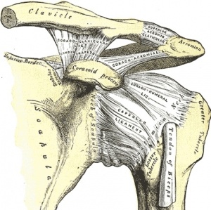
The ACJ capsule and ligaments surrounding the joint work together to provide stability and to keep the clavicle in contact with the acromion process of the scapula.
Joint Capsule: The ACJ has a thin capsule lined with synovium. The capsule is weak and is strengthened by capsular ligaments both inferiorly and superiorly, which in turn is reinforced through attachments from the deltoid ant trapezius.[3] Without the superior and inferior capsular ligaments, the ACJ capsule would not be strong enough to maintain the integrity of the joint.[4]
Ligaments:
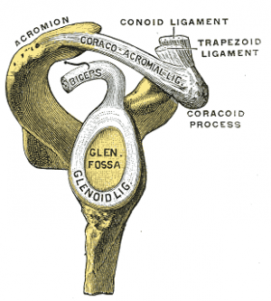
- Coracoclavicular Ligaments[5]: Composed of the conoid and trapezoid ligaments (which do not actually come in contact with the joint), this combined ligament is the primary support ligament of the AC joint. The coracoclavicular ligaments run from the coracoid process to the underside of the clavicle, near the AC joint. These ligaments combribute to horizontal stability, making them crucial for preventing superior dislocation of the AC joint, both portions also limt rotation of the scapula. The most critical roll of the coracoclavicular ligament is in producing the longitudinal rotation of the clavicle necessary for full ROM during elevation of the upper extremity.
- The Conoid Ligament is the fan shaped component of the coracoclavicular ligament. It is located more medially than the trapezoid ligament.
- The Trapezoid Ligamentis the more lateral portion of the coracoclavicular ligament, and is quadrilateral in shape.
- The AC Ligament serves to reinforce the joint capsule and serves as the primary restraint to posterior translation and posterior axial rotation at the AC joint.[6]
Acromioclavicular Joint Disk[edit | edit source]
The disk of the ACJ is variable in size between individuals, at various ages within an individual, and between sides of the same individual. Through 2 years of age, the joint is actually a fibrocar-tilaginous union. With use of the upper extremity, a joint space develops at each articulating surface that may leave a “meniscoid” fibrocartilage remnant within the joint.[2]
Muscles[edit | edit source]
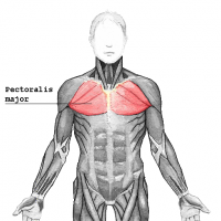
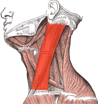
The Clavicle serves as an attachment for many of the muscles that act on the upper extremity and head, these inlude:
- Pectoralis Major (Clavicular head)
- Sternocleidomastoid
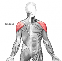
- Deltoid
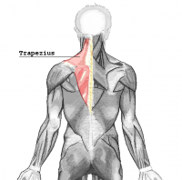
- Trapezius
Motions Available
[edit | edit source]
Motions of the Scapulothoracic joint are generally considered to be a combination of sternoclavicular and acromioclavicular motions.[7] The motions of the ACJ are described as scapular movement with respect to the clavicle, including[8]:
- Upward/downward rotation about an axis directed perpendicular to the scapular plane facing anteriorly and medially.
- Internal/External rotation about an approximately vertical axis.
- Anterior/Posterior tipping or tilting about an axis directed laterally and somewhat anteriorly.
| [9] | [10] |
Closed Packed Position[edit | edit source]
The closed packed position of the ACJ occurs when the GH joint is abducted to 90 degrees.
Open Packed Position[edit | edit source]
The open packed position of the ACJ is undetermined.
Acromioclavicular Stress Tolerance
[edit | edit source]
AC joint is extremely susceptible to both trauma and degenerative change. This is due to its small and incongruent surfaces that result in large forces per unit area. Degenerative change is common from the second decade on, with the joint space itself commonly narrowed by the sixth decade.[2]
Resources[edit | edit source]
- Dutton, M. (2008). Orthopaedic: Examination, evaluation, and intervention (2nd ed.). New York: The McGraw-Hill Companies, Inc.
- Levangie, P.K. & Norkin, C.C. (2005). Joint structure and function: A comprehensive analysis (4th ed.). Philadelphia: The F.A. Davis Company.
Recent Related Research (from Pubmed)[edit | edit source]
Failed to load RSS feed from http://eutils.ncbi.nlm.nih.gov/entrez/eutils/erss.cgi?rss_guid=1FuuOtUS4LZ28aX6oq6sGkDdUT_BXF1bfUXzDcka2jNiMu9Qn|charset=UTF-8|short|max=10: Error parsing XML for RSS
References
[edit | edit source]
- ↑ Dutton, M. (2008). Orthopaedic: Examination, evaluation, and intervention (2nd ed.). New York: The McGraw-Hill Companies, Inc.
- ↑ 2.0 2.1 2.2 2.3 Levangie PK, Norkin CC. Joint Structure and Function : A Comprehensive Analysis. 4th ed. India: JAYPEE; 2006.
- ↑ 3.0 3.1 Neumann DA. Kinesiology of the musculoskeletal system: Foundations for Physical Rehabilitation.2nd Ed.Elsevier Health Sciences;2009
- ↑ Levangie, P.K. and Norkin, C.C. (2005). Joint structure and function: A comprehensive analysis (4th ed.). Philadelphia: The F.A. Davis Company.
- ↑ Levangie, P.K. and Norkin, C.C. (2005). Joint structure and function: A comprehensive analysis (4th ed.). Philadelphia: The F.A. Davis Company.
- ↑ Turnbull, J.R. (1998) Acromioclavicular joint disorders. Med Sci Sports Exerc, 30, S26-32.
- ↑ Inman, V.T., Saudners, J.B., Abbot, L.C. (1996). Observations of the function of the shoulder joint. 1944. Clin Orthop Relat Res. 3-12.
- ↑ Teece, R.M., Lunden, J.B., Lloyd, A.S., Kaiser, A.P., Cieminski, C.J., Ludewig, P.M. (2008). Three-dimensional acromioclavicular motions during elevation of the arm. J Orthop Sports Phys Ther. 38(4), 181-90.
- ↑ Daily Bandha. Shoulder Kinematics. Available from: http://www.youtube.com/watch?v=_Ia0VvT81xc [last accessed 01/12/12]
- ↑ mcp2187. 3D Shoulder. Available from: http://www.youtube.com/watch?v=Jmxz3bEjjGM [last accessed 01/12/12]
