Vagus Nerve
Top Contributors - Kanza Imtiaz, Lucinda hampton and Tony Lowe
This article is currently under review and may not be up to date. Please come back soon to see the finished work! (31/01/2021)
Introduction[edit | edit source]
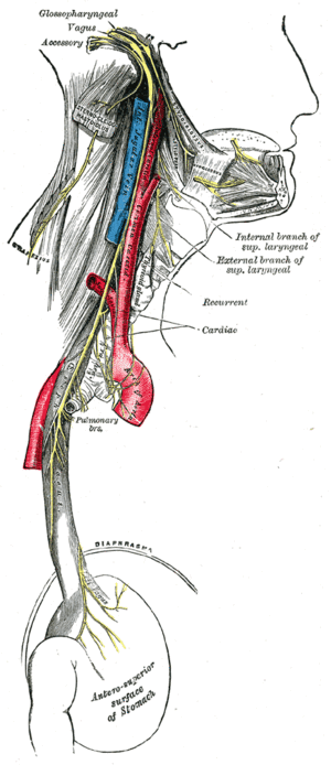
The vagus nerve is the tenth cranial nerve (CN X) and is the longest mixed cranial nerve. The literal translation of the vagus is 'wanderer,' which aptly represents its widespread interfacing of cortex, brainstem, hypothalamus, and the body. Its afferent and efferent pathways comprise about 80% and 20%, respectively[1].
The vagus nerve runs from the brain through the face and thorax to the abdomen. It is a mixed nerve that contains parasympathetic fibres.
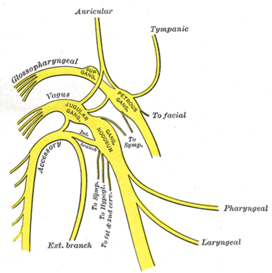
The vagus nerve has two sensory ganglia (masses of nerve tissue that transmit sensory impulses): the superior and the inferior ganglia.
- The branches of the superior ganglion innervate the skin in the concha of the ear.
- The inferior ganglion gives off two branches: the pharyngeal nerve and the superior laryngeal nerve.
The recurrent laryngeal nerve branches from the vagus in the lower neck and upper thorax to innervate the muscles of the larynx (voice box).
The vagus also gives off cardiac, esophageal, and pulmonary branches.
In the abdomen the vagus innervates the greater part of the digestive tract and other abdominal viscera[2].
The accessory nerve (CN XI) joins the vagus nerve just distal to the inferior ganglion.[3]
Structure and Function[edit | edit source]
The vagus nerve contains somatic and visceral afferent fibers, as well as general and special visceral efferent fibers.[4] See Table Below:
| Components | Function | Central component | Cell bodies |
|---|---|---|---|
| Special Visceral Efferent | Swallowing and phonation[4] | Nucleus ambiguus | Nucleus ambiguus |
| General Visceral Efferent | Involuntary muscle control (cardiac, pulmonary, esophageal)
Innervation to glands throughout the gastrointestinal tract[4] |
Dorsal motor nucleus | Dorsal motor nucleus |
| Special Visceral Afferent | Sensations of taste from the tongue and epiglottis [4] | Nucleus tractus solitarius | Inferior ganglion |
| General Visceral Afferent | Sensations from the pharynx, larynx, trachea, esophagus, and the abdominal and thoracic viscera[4] | Nucleus tractus solitarius | Inferior ganglion |
| General Somatic Afferent | Innervation to the external ear and tympanic membrane[4] | Nucleus of the spinal tract of trigeminal | Superior ganglion |
Course[edit | edit source]
The Vagus nerveExits the brain from the medulla oblongata of the brainstem and travels laterally exiting the skull through the jugular foramen.
It descends within the carotid sheath where it is located posterolateral to the internal and common carotid arteries, and medial to the internal jugular vein.
At the base of the neck, the nerve enters the thorax, where the right and left vagus nerve travels on a different path. [7]
- The right vagus enters the thorax by crossing the first part of the subclavian artery and posterior to the innominate artery; then travels behind the primary right bronchus and esophagus to form the esophageal plexus with the left vagus nerve. [8]
- The left vagus enters the thorax by passing between the left common carotid and left subclavian arteries, then travels behind the primary left bronchus and into the esophagus.[8]
Parasympathetic Functions[edit | edit source]
In the thorax and abdomen, the vagus nerve is the main parasympathetic outflow to the heart and gastro-intestinal organs.
The Heart: Cardiac branches arise in the thorax, conveying parasympathetic innervation to the sino-atrial and atrio-ventricular nodes of the heart. These branches stimulate a reduction in the resting heart rate. They are constantly active, producing a rhythm of 60 – 80 beats per minute. If the vagus nerve was lesioned, the resting heart rate would be around 100 beats per minute.
Gastro-Intestinal System: The vagus nerve provides parasympathetic innervation to the majority of the abdominal organs. It sends branches to the oesophagus, stomach and most of the intestinal tract – up to the splenic flexure of the large colon. The function of the vagus nerve is to stimulate smooth muscle contraction and glandular secretions in these organs. For example, in the stomach, the vagus nerve increases the rate of gastric emptying, and stimulates acid production[9].Resources[edit | edit source]
Vagus nerve stimulation
References[edit | edit source]
- ↑ Mandalaneni K, Rayi A. Vagus nerve stimulator. StatPearls [Internet]. 2020 Aug 20.Available from: https://www.ncbi.nlm.nih.gov/books/NBK562175/(accessed 31.1.2021)
- ↑ Britannica Vagus Nerve Available from: https://www.britannica.com/science/vagus-nerve (accessed 31.1.2021)
- ↑ Berthoud HR, Neuhuber WL. Functional and chemical anatomy of the afferent vagal system. Autonomic Neuroscience. 2000 Dec 20;85(1-3):1-7.
- ↑ 4.0 4.1 4.2 4.3 4.4 4.5 Kenny BJ, Bordoni B. Neuroanatomy, Cranial Nerve 10 (Vagus Nerve). InStatPearls [Internet] 2019 Jan 25. StatPearls Publishing.
- ↑ Kenhub - Learn Human Anatomy. Vagus nerve: location, branches and function (preview) - Neuroanatomy | Kenhub. Available from: https://www.youtube.com/watch?v=bNPfjLnnJzA [last accessed 23/9/2020]
- ↑ The Art of Living. What Is The Vagus Nerve? | Vagus Nerve Explained | Brain, Mind Body Connect. Available from: https://www.youtube.com/watch?v=gp67EQhNfj8 [last accessed 23/9/2020]
- ↑ Garner DH, Baker S. Anatomy, Head and Neck, Carotid Sheath. InStatPearls [Internet] 2019 Feb 6. StatPearls Publishing.
- ↑ 8.0 8.1 Yuan H, Silberstein SD. Vagus nerve and vagus nerve stimulation, a comprehensive review: part II. Headache: The Journal of Head and Face Pain. 2016 Feb;56(2):259-66.
- ↑ Teach me anatomy Vagus Nerve Available from: https://teachmeanatomy.info/head/cranial-nerves/vagus-nerve-cn-x/(accessed 31.1.2021)


