Scapholunate Dissociation
Original Editors - Brenda Walk, Liz Record, James Passmore, Jeremy Brady, & Thomas Albaugh as part of the Texas State University Evidence-based Practice Project
Top Contributors - Manisha Shrestha, Kim Jackson, Paul Trevino, Jess Bell and Tarina van der Stockt
Definition/Description[edit | edit source]
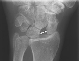
Scapholunate dissociation is the most common and most significant ligamentous injury of the wrist.[1],[2] Scapholunate instability is the most frequent pattern of carpal instability occurring separately and as part of other wrist disorders.[3] It results from the relative instability between the scaphoid and lunate bones secondary to the injury of scapholunate ligament and generally presents radiographically as a widened medial-lateral gap between the two carpal bones.[1]
Anatomy[edit | edit source]
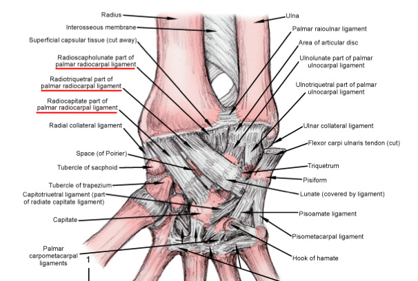
The scapholunate ligament (SLL) also called scapholunate interosseous ligament (SLIL) is C-shaped and has three structurally distinct parts: volar, membranous and dorsal.The dorsal part of the SLL is the strongest and the primary stabiliser of the SL joint and can resist forces of up to 260 N. The avascular proximal membranous portion does not provide any significant laxity restraint (63 N), while the volar part of the SLL (118 N) plays an important role in terms of rotational stability. [3]
The secondary stabilizers are: several extrinsic ligament such as dorsal intercarpal ligament (DIC)[4]
The vascular supply to the scaphoid and SLL is delicate.The main vascular contribution comes from the radial artery.[3]
Epidemiology/Etiology[edit | edit source]
The primary mechanism of injury is an acute stress load of the wrist in extension and ulnar deviation, leading to a force vector pushing the lunate and scaphoid at an angle to each other instead of transferring force directly through the lunate and scaphoid together.[1] This commonly tears the scapholunate ligament and can cause subsequent ligamentous tears of the radiocapitate, radiotriquetral, and dorsal radiocarpal ligaments respectively with increased force loads.
The incidence of scapholunate interosseous ligament injury is unknown due to probable non-diagnosis of this injury in the setting of more distracting injuries resulting from falls on outstretched hands, such as distal radial fractures.[5] Approximately 5% of all wrist sprains have an associated SL tear. With scapholunate disassociations, distal radial fractures (40% of the cases on average), particularly fractures of the radial styloid, the so-called Chauffeur’s fracture and also non-displaced scaphoid fractures are associated.[1][3]
Complications[edit | edit source]
The two primary complications due to scapholunate dissociation are Scapholunate Advanced Collapse (SLAC) and general arthritis of the wrist.[6] Both of these lead to increased disability at the wrist, but a SLAC refers to a specific pattern of osteoarthritis and subluxation resulting secondary to an untreated disassociation that can lead to severe disability at the wrist.
Characteristics/Clinical Presentation[edit | edit source]
Patients will typically present with pain on the radial side of the wrist, specifically localised to the area over the scaphoid and lunate bones. Complaints may include clicking that causes pain, a report of “giving way,” and decreased grip strength. Details to look for in the patient history include injuries to the wrist that involve hyperextension, or repetitive trauma while the wrist is in extension. Lau et al.[7] give an example of prolonged use of crutches as a type of overuse trauma to the wrist while it is in extension.
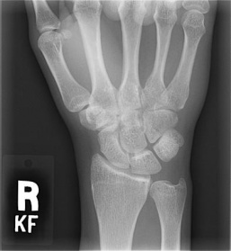
In radiographs, a PA view is used to determine the size of the space between the scaphoid and the lunate. A gap greater than 3mm is considered to be pathologic, although there is research demonstrating that in normal wrists, the average is 3.7mm. For this reason, it is important to compare the size of the gap in the involved wrist to the patient’s uninvolved side. In addition to the PA view, the lateral view shows the scapholunate angle. The normal angle should be 30-60°, and if the angle is over 70°, it is considered a positive scapholunate dissociation.[8]
There are three stages of scapholunate instability. The mildest, occult instability, will typically be due to a FOOSH injury (fall onto outstretched hand), and the patient will have wrist pain, but there will be no visible findings on the radiographs. Watson describes this stage as Pre-Dynamic Instability. The FOOSH injury can cause a tear of the Scapholunate Interosseous Ligament (SLIL) that may or may not progress to a complete tear. The next stage is a complete tear of the SLIL, called Dynamic Scapholunate Instability, and can be diagnosed through imaging.
The next stage, Scapholunate Dissociation, is when not only the SLIL is involved, but also the secondary stabilisers are affected. Eventually, this can lead to degenerative changes, and the patient can develop Scapholunate Advanced Collapse (SLAC),[7] and severe arthrosis of the wrist.
Differential Diagnosis[edit | edit source]
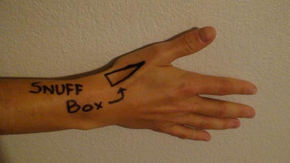
In determining a diagnosis, it is important to take a careful history, including specific details on how the patient fell onto their hand. Falling onto the hand with the wrist extended with ulnar deviation are the most likely positions to lead to damage to the SLIL. During the physical exam, palpation should also be done over the anatomic snuffbox. Tenderness tends to be at the proximal end of the snuffbox, and just distal to Lister’s tubercle. The examiner may also notice a click, or have the patient report the feeling of the wrist “giving way” when pressure is applied over the area. During the acute phase of injury, Scapholunate Dissociation can present similarly to a scaphoid fracture, so it is important that a possible fracture be ruled out. Other possible causes of wrist and hand pain are:
| Site of Pain | Possible Cause |
|---|---|
| Dorsal Wrist |
|
| Ulnar Wrist Pain |
|
| Radial Wrist Pain |
|
| Volar Wrist Pain |
|
| Palmar Pain |
|
Special Radiographic Views for Scapholunate Dissociation: Clenched Fist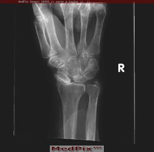
Outcome Measures[edit | edit source]
Examination[edit | edit source]
Signs and Symptoms[edit | edit source]
- Swelling on dorsum of wrist
- Tenderness just distal to Lister's tubercle
- Tenderness in the proximal anatomical snuffbox
- Tenderness at SL joint[11]
- Limited ROM
- Decreased STR
Special Tests[edit | edit source]
- Scapholunate Ballotment Test[11]
- Watson's Test[6][12] Designed as a provocative test with dorsal subluxation of the proximal scaphoid over the dorsal rim of the radius as wrist is radially deviated. To perform the test the examiner:
- Grasps the wrist with their thumb over the patient's scaphoid tuberosity with the wrist in slight dorsiflexion
- Move the patient's wrist from ulnar to radial deviation
- If the scapholunate ligament is disrupted, the scaphoid will tend to flex to the palm of the hand and the lunate will face dorsally on the hand.
- Positive test: The examiner should feel a significant "clunk" and the pt will experience pain.
Used with permission. November 2011. |
Radiographs[edit | edit source]
- AP view
- Lateral view
- Scapholunate gap view[13]
- Clenched fist view (for dynamic wrist instability)[8]
Medical Management[edit | edit source]
The medical management of scapholunate dissociation includes many surgical options, but unfortunately no single procedure has distinguished itself as the best way to manage this injury. Even among surgeons there is no preferred method or current best evidence available. Studies that have attempted to perform a meta-analyses of treatments have found that research articles vary in describing the technical aspects of an operation to reporting the results of using a single method. Evidence based assessments have been attempted, but again, due to the nature of differing methodologies and comparisons of previous reports, no method has been shown as superior. [7][8][8]
While it is difficult for the medical community to decide on specific surgical interventions, other categorical separations do indicate certain procedures over others. These include the acuity or chronicity of the injury, the severity of the separation and to what extent the impairments are affecting the individual.
If the injury is acute, the current methods of choice are:
- Closed reduction and cast immobilisation (8-weeks)
- Closed or open reduction and percutaneous Kershner wire (K-wire) fixation
- Open reduction with direct repair of the scapholunate ligament
- Open reduction and replacement of the scapholunate ligament with tendon graft [7][14]
More often, this type of injury is discovered in the chronic stage as it can present much like a wrist sprain or soft tissue inflammation. Many times, the individual will not even seek care until 15-25 years post injury.[8] With chronic dissociations that present without arthritis, the surgical options are:
- Blatt capsulodesis
- Scaphoid tenodesis
- Tendon reconstruction of the scapholunate ligament
- Scaphoid Trapezium Trapezoid fusion
Other options include (but are much less widely used):
- Arthroscopic joint debridement
- Autogenous bone-retinaculum-bone ligament reconstruction
- Scapholunate or scaphocapitate fusion
If the injury is chronic and has advanced to include arthritic changes (SLAC) options include:
- Proximal carpal row carpectomy
- Scaphoid exision and four-corner fusion of lunate, triquetrum, capitate and hamate
- Wrist fusion[7][15][8]
Soft tissue reconstructions in theory can more accurately restore the normal biomechanics of the wrist. However, the tendons that are used as replacements hold less viscoelasticity than the original ligaments. Skeletal procedures are more predictable, but also more permanent (fusions). Both types of operations have strengths and weaknesses. Garcia-Elias et al. created an algorithm of treatment based off a number of predicating factors of the injury which has been shown to be helpful in deciding which procedure to use. [14]
On a positive note, it has been shown in the literature that surgical management does tend to improve patients’ symptoms. While not always restoring pre-injury function, patients do report better subjective pain ratings and functional outcomes following surgery compared to non-operative treatment.[15] While there are many surgical options, there is no gold standard procedure. Current recommendations suggest that relying on your surgeon’s experience and expertise in a particular method will grant better results that using a standard procedure. [8]
Physical Therapy Management[edit | edit source]
Unfortunately, rehabilitation of SLD has not yet been well researched. As such an impairment based approach is recommended in the acute and chronic phases. It is advised to work closely with the orthopaedic surgeon for post-surgical patients and patients with symptoms that may benefit from surgery.
Acute Phase[edit | edit source]
After ruling out more serious conditions, this condition can be treated as essentially synonymous with an ankle sprain of similar severity.
Chronic Phase[edit | edit source]
Impairments such as limitations in ROM, poor grip strength as compared bilaterally, and severe pain should be addressed as appropriate. It is recommended[16] that the patient should learn to recognise activities which over stress their wrist and avoid or modify these activities. The use of heating or cooling modalities, including contrast baths, and NSAIDS may be used for symptom control during flare-ups.[16] In addition, splints may be used to limit the movement of the involved joints.[16] Cadaver studies have also indicated that the Flexor Carpi Radialis may play a role in stabilising the Scaphoid during movement.[17]
Post-Surgically[edit | edit source]
physiotherapy management should be based on the surgeon's protocol. The patient will likely wear a cast for up to 10 weeks,[18] which can result in a number of significant limitations, which can then be addressed in therapy. It has been suggested by Goldberg et al that rehabilitation following a major reconstruction surgery can take 4-6 months until return to activity and 12-15 months until full recovery.[18] Even with a successful surgery, it is suggested that the patient could benefit from wearing a removable Orthosis during particularly strenuous activities[18].
Effective Exercises[edit | edit source]
- Resisted wrist flexion/extension with a handheld weight. Can also be performed into ulnar/radial deviation or it can be used to stretch the patient into these positions
- Passive self stretch with elbow extended into flexion/extension
- Self resisted isometric strengthening of the wrist extensors/flexors
- Prayer stretch with palms together and fingers extended
- Wrist extension self mobilization in a prayer position
- Concentric/eccentric wrist flexors/extensors resisted by theraband
- Hold a hammer or cane at the end of the handle and move slowly through pronation/supination. As the patient tolerates, move further down the handle to increase the resistance or vice-versa
- Grip strengthening using a hand dynamometer (this can be dosed as a percentage of patients contralateral side or 1 rep max)
Clinical Bottom Line[edit | edit source]
SLD is a very common complication of FOOSH type injuries and is equally commonly undiscovered. There are several secondary complications such as SLAC or arthritis which may not appear until many years after the initial insult. The most well researched treatment option for SLD is surgical repair, but a commonly agreed upon surgical approach has not yet been identified. PT management of the condition should be considered impairment based according to the specific stage and severity of the patient presentation.
References[edit | edit source]
- ↑ 1.0 1.1 1.2 1.3 Duke Orthopaedics: Wheeless' Textbook of Orthopaedics. http://www.wheelessonline.com/ortho/scapholunate_instability (accessed 15 October 2011).
- ↑ Moran SL, Cooney WP, Berger RA, Strickland J. Capsulodesis for the treatment of chronic scapholunate instability. J Hand Surg Am. 2005 Jan;30(1):16-23.
- ↑ 3.0 3.1 3.2 3.3 Andersson JK. Treatment of scapholunate ligament injury: current concepts. EFORT open reviews. 2017 Sep;2(9):382-93.
- ↑ Van Overstraeten L, Camus EJ, Wahegaonkar A, Messina J, Tandara AA, Binder AC, Mathoulin CL. Anatomical description of the dorsal capsulo-scapholunate septum (DCSS)—arthroscopic staging of scapholunate instability after DCSS sectioning. Journal of wrist surgery. 2013 May;2(02):149-54.
- ↑ Tomas A. Scapholunate Dissociation. Journal of Orthopaedic & Sports Physical Therapy. 2018 Mar;48(3):225-.
- ↑ 6.0 6.1 Duke Orthopaedics: Wheeless' Textbook of Orthopaedics. http://www.wheelessonline.com/ortho/scapholunate_advanced_collapse_slac. (Accesed 15 October 2011).
- ↑ 7.0 7.1 7.2 7.3 7.4 Lau S, Swarna SS and Tamvakopoulos GS. Scapholunate dissociation: an overview of the clinical entity and current treatment options. Eur J of Ortho Surgery & Trauma. 2009 Mar;19(6):377-385.
- ↑ 8.0 8.1 8.2 8.3 8.4 8.5 8.6 Bloom HT, Freeland AE, Bowen V, Mrkonjic L. The Treatment of Chronic Scapholunate Dissociation: An Evidence-Based Assessment of the Literature. Orthopedics. 2003;26(2):195-203
- ↑ Jacobson MD, Plancher KD, Evaluation of Hand and Wrist Injuries in Athletes.fckLROperative Techniques in Sports Medicine. 1996; 4(4):210-226
- ↑ J. C. MacDermid, “The patient-rated wrist evaluation (prwe) user manual,”fckLRDecember 2007.
- ↑ 11.0 11.1 Opreanu R, Baulch M, Katranji A. Reduction and maintenance of scapholunate dissociation using the TwinFix screw. Eplasty [serial online]. 2009;9:e7. Available from: MEDLINE with Full Text, Ipswich, MA. http://web.ebscohost.com/ehost/detail?vid=6&hid=19&sid=e64f9dff-7c61-4990-ab69-8ae0a2b413a3%40sessionmgr115&bdata=JnNpdGU9ZWhvc3QtbGl2ZQ%3d%3d Accessed November 26, 2011.
- ↑ Watson's Test. http://en.wikipedia.org/wiki/Watson%27s_test. (accessed 24 October 2011).
- ↑ Akahane M, Ono H, Nakamura T, Kawamura K, Takakura Y. Static scapholunate dissociation diagnosed by scapholunate gap view in wrists with or without distal radius fractures. Hand Surgery: An International Journal Devoted To Hand And Upper Limb Surgery And related Research: Journal Of The Asia-Pacific Federation of Societies For Surgery Of The Hand [serial online]. December 2002;7(2):191-195. Available from: MEDLINE with Full Text, Ipswich, MA. http://web.ebscohost.com/ehost/detail?vid=8&hid=19&sid=e64f9dff-7c61-4990-ab69-8ae0a2b413a3%40sessionmgr115&bdata=JnNpdGU9ZWhvc3QtbGl2ZQ%3d%3d#db=cmedm&AN=12596278 Accessed November 26, 2011.
- ↑ 14.0 14.1 Garcia-Elias M, Lluch AL, Stanley JK. Three-ligament tenodesis for the treatment of scapholunate dissociation: indications and surgical technique. J Hand Surg 2006 Jan;31(1):125–134.
- ↑ 15.0 15.1 Caloia M, Caloia H and Pereira E. Arthroscopic Scapholunate Joint Reduction. Is an Effective Treatment for Irreparable Scapholunate Ligament Tears? Clin Orthop Relat Res. 2011 July. http://www.springerlink.com.libproxy.txstate.edu/content/t748g05668214081/fulltext.pdf Accessed: November 27th, 2011.
- ↑ 16.0 16.1 16.2 Capele A, et al. Mayo Clinic Health Letter - Tools for Healthier Lives. 2011;29(1):1-3. Mayo Foundation for Medical Education and Research, 200 first St. SW, Rochester, MN 55905.http://www.businesswire.com/news/home/20110117005129/en/Mayo-Clinic-Health-Letter-January-2011-Reducing Accessed: November 27th, 2011.
- ↑ Salva-Coll G, et al. The Role of the Flexor Carpi Radialis Muscle in Scapholunate Instability. Jnl Hand Surg – Am Vol. 2011;36A:31-36. Serial online. http://apps.webofknowledge.com.libproxy.txstate.edu/summary.do?SID=4AGo4bcBFcKHjC3AGbl&product=UA&qid=3&search_mode=GeneralSearch Accessed: November 28th, 2011.
- ↑ 18.0 18.1 18.2 Goldberg SH, Strauch RE, Rosenwasser MP. Scapholunate and Lunotriquetral Instability in the Athlete: Diagnosis and Management. Oper Tech Sports Med. 2006;14:108-121. http://www.sciencedirect.com.libproxy.txstate.edu/science/article/pii/S1060187206000402 Accessed: November 27th, 2011.
