When refering to evidence in academic writing, you should always try to reference the primary (original) source. That is usually the journal article where the information was first stated. In most cases Physiopedia articles are a secondary source and so should not be used as references. Physiopedia articles are best used to find the original sources of information (see the references list at the bottom of the article).
If you believe that this Physiopedia article is the primary source for the information you are refering to, you can use the button below to access a related citation statement.
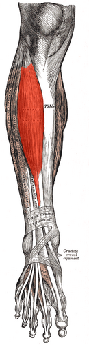
The Tibialis anterior (Tibialis anticus) is situated on the lateral side of the tibia; it is thick and fleshy above, tendinous below. The fibers run vertically downward, and end in a tendon, which is apparent on the anterior surface of the muscle at the lower third of the leg. This muscle overlaps the anterior tibial vessels and deep peroneal nerve in the upper part of the leg.
Variations.—A deep portion of the muscle is rarely inserted into the talus, or a tendinous slip may pass to the head of the first metatarsal bone or the base of the first phalanx of the great toe. The Tibiofascialis anterior, a small muscle from the lower part of the tibia to the transverse or cruciate crural ligaments or deep fascia.[1]
Tibialis anterior is the primary dorsiflexor of the ankle with synergistic action of extensor hallicus longus, extensor digitorium longus and peroneous tertius.
Inversion of the foot.
Adduction of the foot.
Contributor of maintaining the medial arch of the foot.[2]
At anticipatory postural adjustment (APA) phase during gait initiation tibialis anterior favour knee flexion at the stance limb by causing forward displacement of tibia.[3]
Eccentric deceleration of foot plantarflexion, eversion and foot pronation.
Integrated Anatomy
Tibialis anterior is one of the muscles that tend to be inhibited and underactive[4] this leads to overactive of synergistic muscles; extensor hallicus longus, extensor digitorium longus and peroneous tertius.
People with an inhibited or weak Tibialis anterior, for example those with hemiplegia or parkinson's, will have an abnormality in their anticipatory postural adjustment (APA) phase during gait initiation of the affected limb. They will try to compensate for the weakness of Tibialis anterior by overactivity of the contralateral tensor fascia latae (TFL)[3]
Video
Clinical Relevance
Pain along the path of this muscle is often referred to as "Shin splints". Also called medial tibial stress syndrome (MTSS)
Assessment
Palpation
The client is supine. Place your resistance hand on the medial side of the distal foot.
Resist the client from dorsiflexing and inverting the foot. Look the distal tendon of the tibialis anterior on the medial side of the ankle joint and foot; it is usually visible.
Palpate the distal tendon by strumming perpendicular across it. Continue palpating the tibialis anterior proximally to lateral tibial condyle by strumming perpendicular to the fibers.
Once the tibialis anterior has been located, have the client relax it and palpate to asses its baseline tone.[5]
Power
The action of the tibialis anterior muscle is considerably stronger than that of the other three dorsiflexor muscles of the foot.[6]
↑Pasqualino A., Panattoni G.L. 2002 Anatomia Umana. Utet
↑ 3.03.1Jean LH, Marco S, Oscar C , Manh CD. The Neuro-Mechanical Processes That Underlie Goal-Directed Medio-Lateral APA during Gait Initiation. Frontiers Human Neuroscience. August 2016