Thoracolumbar Fascia
Original Editor - Wendy Walker
Lead Editors - Wendy Walker, Lucinda hampton, Kim Jackson, Sehriban Ozmen, WikiSysop, Naomi O'Reilly and Bruno Serra
Description[edit | edit source]
The thoracolumbar fascia is a large area of connective tissue - roughly diamond-shaped - which comprises the thoracic and lumbar parts of the deep fascia enclosing the intrinsic back muscles.
Most developed in the lumbar region, it consists of multiple layers of crosshatched collagen fibres that cover the back muscles in the lower thoracic and lumbar area before passing through these muscles to attach to the sacral bone.
Anatomy 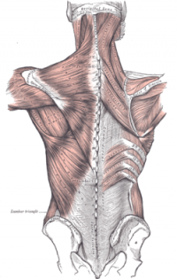 [edit | edit source]

The TLF is a complex of several layers that separates the paraspinal muscles from the muscles of the posterior abdominal wall, quadratus lumborum (QL) and psoas major[1].
Numerous descriptions of this structure have presented either a two-layered model or a three-layered model.
In the thoracic region, it forms a thin covering for the extensor muscles of the vertebral column. Medially, it is attached to the spines of the thoracic vertebrae, and laterally it is attached to the ribs, near their angles. In the lumbar region, it is also attached to the vertebral spines, but in addition it forms a strong aponeurosis that is connected laterally to the flat muscles of the abdominal wall. Medially, it splits into anterior, middle and posterior layers. The first two layers surround quadratus lumborum and the last two form a sheath for the erector spinae and multifidus muscles. Below, it is attached to the iliolumbar ligament, the iliac crest and the sacroiliac joint. Via its extensive attachment to vertebral spines, the thoracolumbar fascia is attached to the supraspinous and interspinous ligaments and to the capsule of the facet joints.
The illustration opposite shows the superficial muscles of the back. The thoracolumbar fascia is the gray area at bottom centre.
The superficial lamina of the posterior layer of the TLF (PLF) is dominated by the aponeuroses of the latissimus dorsi and the serratus posterior inferior. The deeper lamina of the PLF forms an encapsulating retinacular sheath around the paraspinal muscles.
The middle layer of the TLF (MLF) appears to derive from an intermuscular septum that developmentally separates the epaxial from the hypaxialmusculature.
Function[edit | edit source]
The unyielding character of the deep fascia enables it to serve as a means of containing and separating groups of muscles into relatively well-defined spaces called ‘compartments’. The deep fascia integrates these compartments and transmits load between them.
The TLF is a critical part of a myofascial girdle that surrounds the lower portion of the torso, playing an important role in the following functions[1]:
- posture
- load transfer
- respiration
Load Transfer between upper and lower limbs
[edit | edit source]
Vleeming et al[2] have highlighted the importance of the thoracolumbar fascia in integrating the activity of muscles traditionally regarded as belonging to the lower limb, upper limb, spine or pelvis and whose action is thus often considered in that territory alone. They have argued that a common attachment to the thoracolumbar fascia means that the latter has an important role in integrating load transfer between different regions. In particular, these authors have proposed that gluteus maximus and latissimus dorsi (two of the largest muscles of the body) contribute to co-ordinating the contralateral pendulum like motions of the upper and lower limbs that characterize running or swimming[2]. They suggest that the muscles do so because of a shared attachment to the posterior layer of the thoracolumbar fascia.
Pathology/Injury[edit | edit source]
Techniques[edit | edit source]
Palpation[edit | edit source]
Examination[edit | edit source]
Treatment[edit | edit source]
Resources[edit | edit source]
Recent Related Research (from Pubmed)[edit | edit source]
Extension:RSS -- Error: Not a valid URL: Feed goes here!!|charset=UTF-8|short|max=10
References[edit | edit source]
References will automatically be added here, see adding references tutorial.
- ↑ 1.0 1.1 Cite error: Invalid
<ref>tag; no text was provided for refs namedWillard et al - ↑ 2.0 2.1 The posterior layer of the thoracolumbar fascia. Its function in load transfer from spine to legs.fckLRVleeming A, Pool-Goudzwaard AL, Stoeckart R, van Wingerden JP, Snijders CJfckLRfckLRSpine (Phila Pa 1976). 1995 Apr 1; 20(7):753-8.

