Search results
Page title matches
- Previous literature supports the use of Tai Chi, an ancient Chinese exercise focusing on relaxation techniques, ...ible within the UK, like Yoga, Pilates and Tai Chi. The study also did not use a secondary follow-up period, which questions the validity and longevity of46 KB (6,543 words) - 16:21, 11 October 2023
Page text matches
File:Sagittal section of the knee joint Primal.png ...ven permission to use this image exclusively in Physiopedia. Please do not use this image outside Physiopedia unless you have prior permission(660 × 660 (607 KB)) - 21:46, 7 December 2020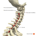
File:Muscles of the cervical region multifidus deep layer Primal.png ...ven permission to use this image exclusively in Physiopedia. Please do not use this image outside Physiopedia unless you have prior permission(660 × 660 (290 KB)) - 15:17, 8 December 2020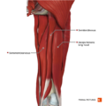
File:Muscles of the thigh posterior compartment Primal.png ...ven permission to use this image exclusively in Physiopedia. Please do not use this image outside Physiopedia unless you have prior permission(660 × 660 (315 KB)) - 20:49, 7 December 2020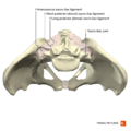
File:Sacro-iliac joint Primal.png ...ven permission to use this image exclusively in Physiopedia. Please do not use this image outside Physiopedia unless you have prior permission(660 × 660 (239 KB)) - 00:49, 8 December 2020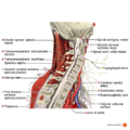
File:Sagittal section of the cervical spine Primal.png ...ven permission to use this image exclusively in Physiopedia. Please do not use this image outside Physiopedia unless you have prior permission(990 × 990 (792 KB)) - 15:39, 8 December 2020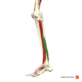
File:Peroneus brevis Primal.png ...ven permission to use this image exclusively in Physiopedia. Please do not use this image outside Physiopedia unless you have prior permission(660 × 660 (113 KB)) - 15:15, 10 January 2021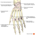
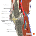
File:Ligaments of the hand dorsal aspect Primal.png ...ven permission to use this image exclusively in Physiopedia. Please do not use this image outside Physiopedia unless you have prior permission(660 × 660 (297 KB)) - 00:20, 7 December 2020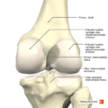
File:Knee joint posterior aspect Primal.png ...ven permission to use this image exclusively in Physiopedia. Please do not use this image outside Physiopedia unless you have prior permission(660 × 660 (289 KB)) - 21:37, 7 December 2020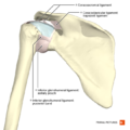
File:Ligaments of the shoulder posterior aspect Primal.png ...ven permission to use this image exclusively in Physiopedia. Please do not use this image outside Physiopedia unless you have prior permission(660 × 660 (256 KB)) - 01:06, 8 December 2020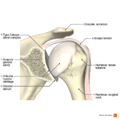
File:Illustration of type I biceps labral complex Primal.png ...ven permission to use this image exclusively in Physiopedia. Please do not use this image outside Physiopedia unless you have prior permission(990 × 990 (498 KB)) - 01:13, 8 December 2020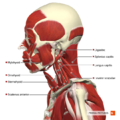
File:Intermediate muscles of the head and neck lateral aspect Primal.png ...ven permission to use this image exclusively in Physiopedia. Please do not use this image outside Physiopedia unless you have prior permission(660 × 660 (437 KB)) - 20:06, 7 December 2020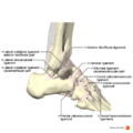
File:Ligaments of the ankle lateral aspect Primal.png ...ven permission to use this image exclusively in Physiopedia. Please do not use this image outside Physiopedia unless you have prior permission(990 × 990 (433 KB)) - 00:16, 8 December 2020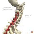
File:Muscles of the cervical region multifidus intermediate layer Primal.png ...ven permission to use this image exclusively in Physiopedia. Please do not use this image outside Physiopedia unless you have prior permission(660 × 660 (307 KB)) - 15:18, 8 December 2020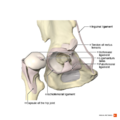
File:Hip joint Primal.png ...ven permission to use this image exclusively in Physiopedia. Please do not use this image outside Physiopedia unless you have prior permission(990 × 990 (443 KB)) - 20:52, 7 December 2020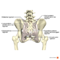
File:Ligaments of the pelvis posterior aspect Primal.png ...ven permission to use this image exclusively in Physiopedia. Please do not use this image outside Physiopedia unless you have prior permission(990 × 990 (537 KB)) - 00:50, 8 December 2020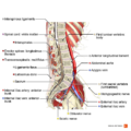
File:Sagittal section of the lumbar spine Primal.png ...ven permission to use this image exclusively in Physiopedia. Please do not use this image outside Physiopedia unless you have prior permission(990 × 990 (683 KB)) - 15:41, 8 December 2020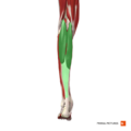
File:Gastrocnemius Primal.png ...ven permission to use this image exclusively in Physiopedia. Please do not use this image outside Physiopedia unless you have prior permission(660 × 660 (112 KB)) - 15:16, 10 January 2021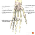
File:Ligaments of the hand palmar aspect Primal.png ...ven permission to use this image exclusively in Physiopedia. Please do not use this image outside Physiopedia unless you have prior permission(660 × 660 (288 KB)) - 00:22, 7 December 2020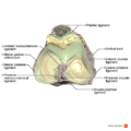
File:Ligaments of the knee joint superior aspect Primal.png ...ven permission to use this image exclusively in Physiopedia. Please do not use this image outside Physiopedia unless you have prior permission(990 × 990 (409 KB)) - 21:37, 7 December 2020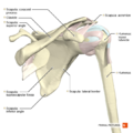
File:Shoulder anterior aspect Primal.png ...ven permission to use this image exclusively in Physiopedia. Please do not use this image outside Physiopedia unless you have prior permission(660 × 660 (266 KB)) - 01:07, 8 December 2020


